
Page 292 - Atlas of Histology with Functional Correlations
P. 292
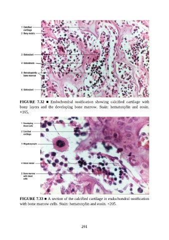
FIGURE  7.32  ■  Endochondral  ossification  showing  calcified  cartilage  with
               bony  layers  and  the  developing  bone  marrow.  Stain:  hematoxylin  and  eosin.
               ×165.


               FIGURE 7.33 ■ A section of the calcified cartilage in endochondral ossification

               with bone marrow cells. Stain: hematoxylin and eosin. ×205.


                                                          291