Page 738 - Atlas of Histology with Functional Correlations
P. 738
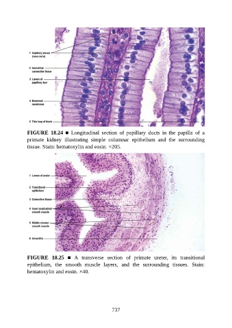
FIGURE  18.24  ■  Longitudinal  section  of  papillary  ducts  in  the  papilla  of  a
               primate  kidney  illustrating  simple  columnar  epithelium  and  the  surrounding
               tissue. Stain: hematoxylin and eosin. ×205.


               FIGURE  18.25  ■  A  transverse  section  of  primate  ureter,  its  transitional
               epithelium,  the  smooth  muscle  layers,  and  the  surrounding  tissues.  Stain:
               hematoxylin and eosin. ×40.


                                                          737

