
Page 952 - Atlas of Histology with Functional Correlations
P. 952
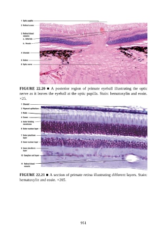
FIGURE  22.20  ■  A  posterior  region  of  primate  eyeball  illustrating  the  optic
               nerve as it leaves the eyeball at the optic papilla. Stain: hematoxylin and eosin.
               ×25.


               FIGURE 22.21 ■ A section of primate retina illustrating different layers. Stain:

               hematoxylin and eosin. ×205.


                                                          951