
Page 488 - AWSAR 2.0
P. 488
464 || AWSAR Awarded Popular Science Stories - 2019
(c) production of new proteins or even (d) migration of the whole cell. But, how does the cellmembranecontroltheformationofspecific protein-lipid assemblies in the right place and at the right time?
The cell membrane is draped on a specialized actin network called actin cortex. Actin is a small, round protein and can exist as individual units or filaments of variable length. The composition and architecture of the actin cortex are regulated by actin modulators and myosin motors. Actin modulators can polymerize, depolymerize, break and bundle actin filaments. Myosin motors are specialized proteins that convert the chemical energy of ATP (the energy currency of a cell) into mechanical energy. Many myosin molecules can intertwine to form thread-like structures called myofilaments. These myofilaments can simultaneously walk on multiple actin filaments (or bundles) and induce long-range movements or flows in the resulting
actomyosin aggregates (e.g., during muscle contraction). If these actomyosin flows generated on the cortex can be coupled to cell membrane components, its implications in membrane organization can be tremendous.
Various models have
been proposed to explain the
cell membrane organization.
Few models propose that
certain membrane lipids tend
to stick together due to like-
like interactions and organize
into membrane patches.
Specific proteins can prefer or avoid these lipid patches, which can be a mechanism tosortandlocalizemembranecomponents (lipid raft model). Other models suggest that transmembrane proteins, which can directly bind to the actin cortex, create fences along
the actin mesh and hinder long-range mobility of other membrane components (picket-fence model). Also, there are other models that essentially propose a combination of both these mechanisms. While these models have successfully explained the organization of a range of membrane components, there is a growing class of phenomena that appears inconsistent with these models and hence merits a fulfilling explanation.
Recent studies on animal cells grown under controlled conditions in laboratories across the globe have revealed a more engaging role of the actin cortex in cell membrane organization. Several membrane proteins bind to actin filaments, either directly through their actin-binding domains or indirectly via adaptor molecules. Many of these membrane proteins have been shown to exist in tiny, transient and localized clusters driven by actomyosin flows generated on the cortex. These results
have suggested that the cell surface behaves more like a composite of the membrane and the actin cortex. This has been suggested as a potential mechanism for the local control of plasma membrane composition.
This active composite membrane model, as it’s called, posits that the actin cortex comprises short actin filaments along with relatively stable actin bundles. These short filaments can rapidly grow on one end and shrink on the other, and can transiently attach to the
cell membrane via linker proteins (membrane proteins that can bind to actin filaments and create membrane–cortex connections). Myosin motors act on these dynamic actin filaments and produce ATP-fuelled contractile flows in these actomyosin complexes, leading
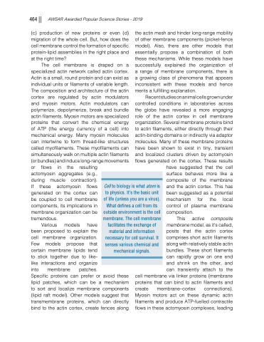
Cell to biology is what atom is to physics. It’s the basic unit of life (unless you are a virus). What defines a cell from its outside environment is the cell membrane. The cell membrane facilitates the exchange of material and information necessary for cell survival. It senses various chemical and mechanical signals.
