Page 2 - Urinary Incontinence
P. 2
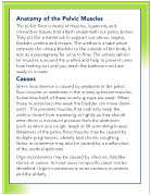
Anatomy of the Pelvic Muscles
The pelvic floor consists of muscles, ligaments and
connective tissues that attach underneath our pelvic bones.
They act like a hammock to support our uterus, vagina,
bladder, urethra and rectum. The urethra is a tube which
connects the urinary bladder to the outside of the body. It
acts as a passageway for urine to flow. The urinary sphinc-
ter muscles surround the urethra and help to prevent urine
from leaking out until you reach the bathroom and are
ready to urinate.
Causes
Stress incontinence is caused by weakness in the pelvic
floor muscles or weakness in the urinary sphincter muscles.
Sometimes both of these muscle groups are weak. When
these muscles become weak the bladder can move down-
ward . This prevents muscles that ordinarily keep the
urethra closed from squeezing as tightly as they should
when there is increased pressure from the abdomen
(such as when you cough, laugh or lift something heavy).
Weakness of the pelvic floor muscles may be caused by
multiple pregnancies, obesity and chronic coughing.
Stress incontinence may also be caused by a malfunction
of the urethral sphincter.
Urge incontinence may be caused by infection, bladder
stones or cancer. In most cases no specific cause can be
identified. Urge incontinence is most common in women
and the elderly.