
Page 10 - DMX HANDBOOK 4TH EDITION
P. 10
8. Ligament injuries produce two types of “motion segment” impairment – translational and angular.
The primary function of ligaments is to hold the vertebrae in place.
Ligamentous “stretch” injuries produce laxity. Ligamentous laxity, in turn, causes abnormal movement of the vertebrae that can be
seen on X-ray. The ligaments do not show up on the DMX. However, the abnormal positioning and motion of the vertebrae in the
cervical spine does show up on a motion X-ray. It is a simple fact that the function of ligaments is to keep the vertebrae in their
normal anatomical position. Abnormal positioning of the vertebrae results from ligamentous laxity. Therefore, the abnormal motion
of the vertebrae revealed by motion X-ray proves the damage of the ligaments, i.e., ligamentous laxity.
Medical literature has established two types of motion segment impairment resulting from ligamentous laxity:
1. Translational irregularity,
and
2. Angular irregularity.
Since 1993, the AMA Guides to the Evaluation of Permanent Impairment have recognized the concept of “Alteration of Motion
Segment Impairment” (AOMSI).
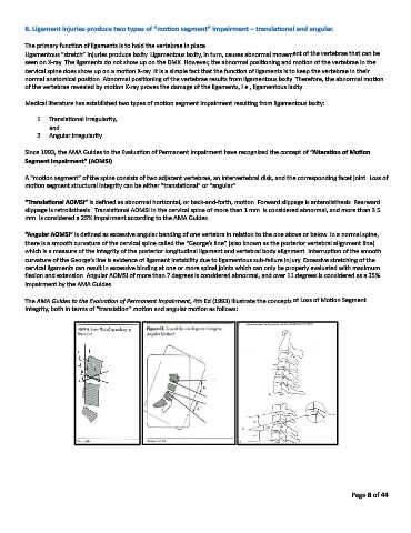
A “motion segment” of the spine consists of two adjacent vertebrae, an intervertebral disk, and the corresponding facet joint. Loss of
motion segment structural integrity can be either “translational” or “angular”.
“Translational AOMSI” is defined as abnormal horizontal, or back-and-forth, motion. Forward slippage is anterolisthesis. Rearward
slippage is retrolisthesis. Translational AOMSI in the cervical spine of more than 1 mm. is considered abnormal, and more than 3.5
mm. is considered a 25% impairment according to the AMA Guides.
“Angular AOMSI” is defined as excessive angular bending of one vertebra in relation to the one above or below. In a normal spine,
there is a smooth curvature of the cervical spine called the “George’s line” (also known as the posterior vertebral alignment line)
which is a measure of the integrity of the posterior longitudinal ligament and vertebral body alignment. Interruption of the smooth
curvature of the George’s line is evidence of ligament instability due to ligamentous sub-failure injury. Excessive stretching of the
cervical ligaments can result in excessive binding at one or more spinal joints which can only be properly evaluated with maximum
flexion and extension. Angular AOMSI of more than 7 degrees is considered abnormal, and over 11 degrees is considered as a 25%
impairment by the AMA Guides.
The AMA Guides to the Evaluation of Permanent Impairment, 4th Ed (1993) illustrate the concepts of Loss of Motion Segment
Integrity, both in terms of “translation” motion and angular motion as follows:
Page 8 of 44