
Page 119 - MNU-PM503 Parasitology practical book
P. 119
Pharm D- Clinical Pharmacy Program Third Level Parasitology and virology (PM501)
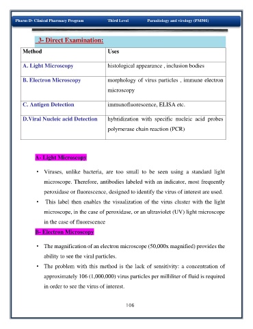
3- Direct Examination:
Method Uses
o
g
o
l
t
s
i
h
ca
i
A. Light Microscopy l appearance , inclusion bodies
B. Electron Microscopy morphology of virus particles , immune electron
microscopy
C. Antigen Detection immunofluorescence, ELISA etc.
D.Viral Nucleic acid Detection hybridization with specific nucleic acid probes
polymerase chain reaction (PCR)
A- Light Microscopy
• Viruses, unlike bacteria, are too small to be seen using a standard light
microscope. Therefore, antibodies labeled with an indicator, most frequently
peroxidase or fluorescence, designed to identify the virus of interest are used.
• This label then enables the visualization of the virus cluster with the light
microscope, in the case of peroxidase, or an ultraviolet (UV) light microscope
in the case of fluorescence
B- Electron Microscopy
• The magnification of an electron microscope (50,000x magnified) provides the
ability to see the viral particles.
• The problem with this method is the lack of sensitivity: a concentration of
approximately 106 (1,000,000) virus particles per milliliter of fluid is required
in order to see the virus of interest.
106