
Page 22 - DP Vol 19 No 6 pw_Neat
P. 22
RestoRative section
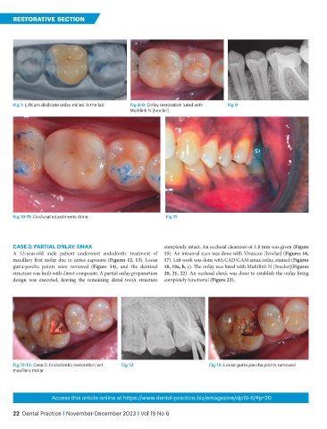
Fig 7: Lithium disilicate onlay milled in the lab Fig 8-9: Onlay restoration luted with Fig 9
Multilink N [Ivoclar]
Fig 10-11: Occlusal adjustments done Fig 11
CASE 2: PARTIAL ONLAY: EMAX completely intact. An occlusal clearance of 1.8 mm was given (Figure
A 52-year-old male patient underwent endodontic treatment of 15). An intraoral scan was done with Vivascan [Ivoclar] (Figures 16,
maxillary first molar due to caries exposure (Figures 12, 13). Loose 17). Lab work was done with CAD/CAM emax onlay, stained (Figures
gutta-percha points were removed (Figure 14), and the dentinal 18, 19a, b, c). The onlay was luted with Multilink N [Ivoclar](Figures
structure was built with direct composite. A partial onlay preparartion 20, 21, 22). An occlusal check was done to establish the onlay being
design was executed, leaving the remaining distal tooth structure completely functional (Figure 23).
Fig 12-13: Case 2: Endodontic restoration wrt Fig 13 Fig 14: Loose gutta-percha points removed
maxillary molar
Access this article online at https://www.dental-practice.biz/emagazine/dp19-6/#p=20
22 Dental Practice I November-December 2023 I Vol 19 No 6