
Page 61 - DP Vol 19 No 6 pw_Neat
P. 61
1c
1a 1b
Fig 1a, b & c: Pre-operative extraoral and intraoral images
3c
3a 3b 3d
Fig 2: Pre-operative OPG Fig 3a-d: Digital scanning of maxillary and mandibular arches
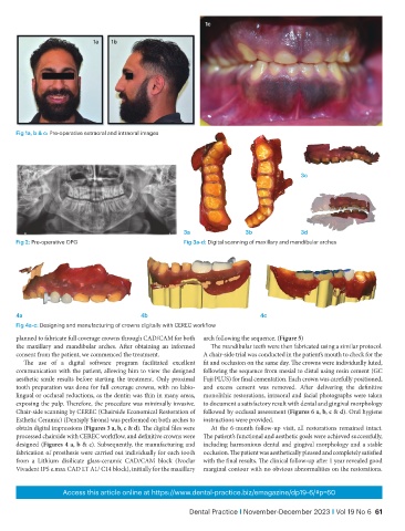
4a 4b 4c
Fig 4a-c: Designing and manufacturing of crowns digitally with CEREC workflow
planned to fabricate full coverage crowns through CAD/CAM for both arch following the sequence. (Figure 5)
the maxillary and mandibular arches. After obtaining an informed The mandibular teeth were then fabricated using a similar protocol.
consent from the patient, we commenced the treatment. A chair-side trial was conducted in the patient’s mouth to check for the
The use of a digital software program facilitated excellent fit and occlusion on the same day. The crowns were individually luted,
communication with the patient, allowing him to view the designed following the sequence from mesial to distal using resin cement (GC
aesthetic smile results before starting the treatment. Only proximal Fuji PLUS) for final cementation. Each crown was carefully positioned,
tooth preparation was done for full coverage crowns, with no labio- and excess cement was removed. After delivering the definitive
lingual or occlusal reductions, as the dentin was thin in many areas, monolithic restorations, intraoral and facial photographs were taken
exposing the pulp. Therefore, the procedure was minimally invasive. to document a satisfactory result with dental and gingival morphology
Chair-side scanning by CEREC (Chairside Economical Restoration of followed by occlusal assessment (Figures 6 a, b, c & d). Oral hygiene
Esthetic Ceramic) (Dentsply Sirona) was performed on both arches to instructions were provided.
obtain digital impressions (Figures 3 a, b, c & d). The digital files were At the 6-month follow-up visit, all restorations remained intact.
processed chairside with CEREC workflow, and definitive crowns were The patient’s functional and aesthetic goals were achieved successfully,
designed (Figures 4 a, b & c). Subsequently, the manufacturing and including harmonious dental and gingival morphology and a stable
fabrication of prosthesis were carried out individually for each tooth occlusion. The patient was aesthetically pleased and completely satisfied
from a Lithium disilicate glass-ceramic CAD/CAM block (Ivoclar with the final results. The clinical follow-up after 1 year revealed good
Vivadent IPS e.max CAD LT A1/ C14 block), initially for the maxillary marginal contour with no obvious abnormalities on the restorations.
Access this article online at https://www.dental-practice.biz/emagazine/dp19-6/#p=60
Dental Practice i November-December 2023 i Vol 19 No 6 61