
Page 37 - Dental Practice Vol 17 No.5_
P. 37
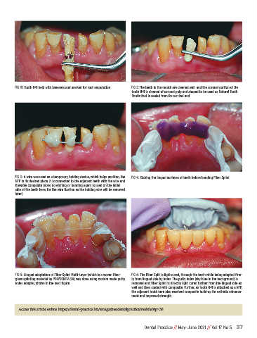
FIG 1B: Tooth #41 held with tweezers and marked for root amputation  FIG 2: The teeth in the mouth are cleaned well and the coronal portion of the
                                                                   tooth #41 is cleaned of coronal pulp and shaped to be used as Natural Tooth
                                                                   Pontic that is sealed from its cervical end.


              FIG 3: A wire was used as a temporary holding device, which helps position, the  FIG 4: Etching the lingual surfaces of teeth before bonding Fiber Splint
              NTP in its desired place. It is connected to the adjacent teeth with the wire and
              flowable composite (note: no etching or bonding agent is used on the labial
              side of the teeth here, for the wire fixation as the holding wire will be removed
              later)


              FIG 5: Lingual adaptation of Fiber Splint Multi-Layer (which is a woven fiber-  FIG 6: The Fiber Split is light cured, through the teeth while being adapted firm-
              glass splinting material by POLYDENTIA SA) was done using custom made putty  ly from lingual side by index. The putty index (sky-blue in the background) is
              index adapter, shown in the next figure.             removed and Fiber Splint is directly light cured further from the lingual side as
                                                                   well and then coated with composite. Further, as tooth #41 is attached as a NTP,
                                                                   the adjacent tooth here also received composite build-up for esthetic enhance-
                                                                   ment and improved strength.


               Access this article online https://dental-practice.biz/emagazine/dentalpractice/mobile/#p=36


                                                                         Dental Practice // May-June 2021 // Vol 17 No 5 37