
Page 525 - Physics Coursebook 2015 (A level)
P. 525
Chapter 32: Medical imaging
hard X-rays. In contrast, investigation of the tissue of the breast, where the tissue is a poor absorber, will require a longer exposure, using much softer (long-wavelength, low- frequency) X-rays.
As we have seen, different tissues show up differently in X-ray images. In particular, bone can readily be distinguished from soft tissue such as muscle because it is a good absorber of X-rays. However, it is often desirable to show up different soft tissues that absorb X-rays equally. In order to do this, contrast media are used.
A contrast medium is a substance such as iodine or barium which is a good absorber of X-rays. The patient may swallow a barium-containing liquid (a ‘barium meal’), or have a similar liquid injected into the tissue of interest. This tissue is then a better absorber of X-rays and its edges show up more clearly on the final image.
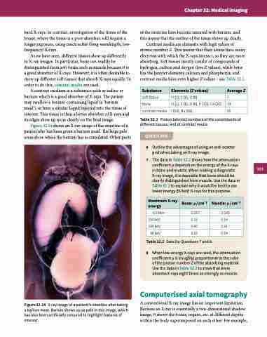
Figure 32.14 shows an X-ray image of the intestine of a patient who has been given a barium meal. The large pale areas show where the barium has accumulated. Other parts
Figure 32.14 X-ray image of a patient’s intestine after taking a barium meal. Barium shows up as pale in this image, which has also been artificially coloured to highlight features of interest.
of the intestine have become smeared with barium, and this means that the outline of the tissue shows up clearly.
Contrast media are elements with high values of atomic number Z. This means that their atoms have many electrons with which the X-rays interact, so they are more absorbing. Soft tissues mostly consist of compounds of hydrogen, carbon and oxygen (low Z values), while bone has the heavier elements calcium and phosphorus, and contrast media have even higher Z values – see Table 32.1.
Substance
Elements (Z values)
Average Z
soft tissue
bone
contrast media
H (1), C (6), O (8) 7
H (1), C (6), O (8), P (15), Ca (20) 14
I (53), Ba (56) 55
Table 32.1 Proton (atomic) numbers of the constituents of different tissues, and of contrast media.
QUESTIONS
6 Outline the advantages of using an anti-scatter grid when taking an X-ray image.
7 The data in Table 32.2 shows how the attenuation coefficient μ depends on the energy of the X-rays in bone and muscle. When making a diagnostic X-ray image, it is desirable that bone should be clearly distinguished from muscle. Use the data in Table 32.2 to explain why it would be best to use lower energy (50 keV) X-rays for this purpose.
Table 32.2 Data for Questions 7 and 8.
8 When low-energy X-rays are used, the attenuation coefficient μ is (roughly) proportional to the cube of the proton number Z of the absorbing material. Use the data in Table 32.2 to show that bone absorbs X-rays eight times as strongly as muscle.
Computerised axial tomography
A conventional X-ray image has an important limitation. Because an X-ray is essentially a two-dimensional shadow image, it shows the bones, organs, etc. at different depths within the body superimposed on each other. For example,
Maximum X-ray energy
Bone: μ / cm−1
Muscle: μ / cm−1
4.0 MeV
0.087
0.049
250 keV
0.32
0.16
100 keV
0.60
0.21
50 keV
3.32
0.54
513
