
Page 526 - Physics Coursebook 2015 (A level)
P. 526
Cambridge International A Level Physics
514
in Figure 32.15, it is difficult to distinguish the bones of the front and back of the ribcage. This can be overcome by taking several images at different angles. An experienced radiographer can then study these images and deduce what is going on inside the patient.
Figure 32.15 Computer-generated X-ray image of a person in a yoga position. This shows the difficulty of distinguishing one bone from another when they overlap.
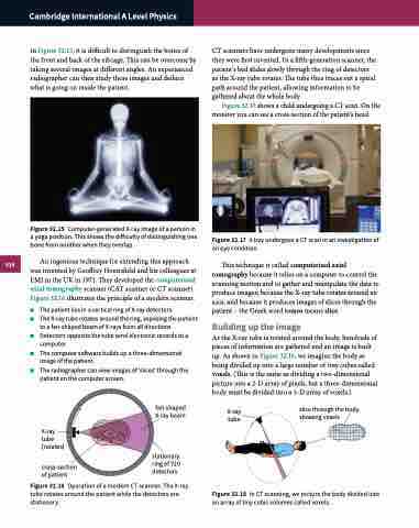
An ingenious technique for extending this approach was invented by Geoffrey Hounsfield and his colleagues at EMI in the UK in 1971. They developed the computerised axial tomography scanner (CAT scanner or CT scanner). Figure 32.16 illustrates the principle of a modern scanner.
■■ The patient lies in a vertical ring of X-ray detectors.
■■ The X-ray tube rotates around the ring, exposing the patient
to a fan-shaped beam of X-rays from all directions.
■■ Detectors opposite the tube send electronic records to a
computer.
■■ The computer software builds up a three-dimensional
image of the patient.
■■ The radiographer can view images of ‘slices’ through the
CT scanners have undergone many developments since they were first invented. In a fifth-generation scanner, the patient’s bed slides slowly through the ring of detectors as the X-ray tube rotates. The tube thus traces out a spiral path around the patient, allowing information to be gathered about the whole body.
Figure 32.17 shows a child undergoing a CT scan. On the monitor you can see a cross-section of the patient’s head.
Figure 32.17 A boy undergoes a CT scan in an investigation of an eye condition.
This technique is called computerised axial tomography because it relies on a computer to control the scanning motion and to gather and manipulate the data to produce images; because the X-ray tube rotates around an axis; and because it produces images of slices through the patient – the Greek word tomos means slice.
Building up the image
As the X-ray tube is rotated around the body, hundreds of pieces of information are gathered and an image is built up. As shown in Figure 32.18, we imagine the body as being divided up into a large number of tiny cubes called voxels. (This is the same as dividing a two-dimensional picture into a 2-D array of pixels, but a three-dimensional body must be divided into a 3-D array of voxels.)
patient on the computer screen.
X-ray tube (rotates)
cross-section of patient
fan-shaped X-ray beam
stationary ring of 720 detectors
X-ray tube
slice through the body showing voxels
Figure 32.16 Operation of a modern CT scanner. The X-ray tube rotates around the patient while the detectors are stationary.
Figure 32.18 In CT scanning, we picture the body divided into an array of tiny cubic volumes called voxels.
