
Page 528 - Physics Coursebook 2015 (A level)
P. 528
Cambridge International A Level Physics
516
Steps 3 and 4
The beam is rotated twice more through 45° and each time the detected values are added to the memory grid.
Step 5
Now each voxel in the array has been exposed to X-rays from four different directions. How can we extract the original values from the final memory grid? Note that, in each step, the total density detected had a value of
21 (10+11 in Step 1, 5+8+8 in Step 2, and so on). We subtract this background value from each square in the memory grid, and then divide the remainder by three. The final values in the memory grid are the same as in the original 2 × 2 array.
Our 2 × 2 array is an example in two dimensions. In three dimensions, we would need to consider a 2 × 2 × 2 array
of cubes rather than squares. This is known as an 8-voxel cube. A real object would be made up of a very large number of tiny voxels in three dimensions, requiring very powerful computers to analyse all the data collected.
For a well-defined image in a CT scan, we need the voxels to be small. Two things are needed to achieve this:
■■ The X-ray beam must be well collimated so that it consists of parallel rays – the rays must not spread outwards.
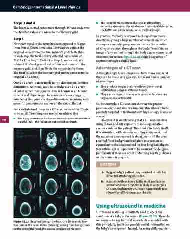
Figure 32.20 Sections through the head of a 10-year-old boy. You can see the haematoma (bruising) arising from being struck on the side of the head; this causes pressure on his brain.
■■ The detector must consist of a regular array of tiny detecting elements – the smaller each individual detector is, the better will be the resolution in the final image.
In practice, the body is exposed to X-rays from many directions, giving a large number of values from which
a complex computer program can deduce the variation
of X-ray absorption throughout the body. From this, an image of any section through the body can be constructed on a monitor screen. Figure 32.20 shows a sequence of sections through a child’s head.
Advantages of a CT scan
Although single X-ray images still have many uses (and they can be made very quickly), CT scans have a number of advantages:
■■ They produce images that show three-dimensional relationships between different tissues.
■■ They can distinguish tissues with quite similar densities (attenuation coefficients).
So, for example, a CT scan can show up the precise position, shape and size of a tumour. This allows it to be precisely targeted in treatment with high-energy X-rays or γ-rays.
However, it is worth noting that a CT scan involves using X-rays and any exposure to ionising radiation carries a risk for the patient. These risks are fairly small; it is estimated, with modern scanning equipment, that the radiation dose received is about one-third the dose received from background radiation in a year, or is equivalent to the dose received on four long-haul flights. Nevertheless, it is important to be aware of the dangers, particularly if there are other underlying health problems or if a woman is pregnant.
QUESTIONS
9 Suggest why a patient may be asked to hold his or her breath during a CT scan.
10 A patient with an injury to the skull, perhaps as a result of a road accident, is likely to undergo a CT scan. Explain why a CT scan is preferable to a conventional X-ray in a case like this.
Using ultrasound in medicine
Ultrasound scanning is routinely used to check the condition of a baby in the womb (Figure 32.21). There do not seem to be any harmful side-effects associated with this procedure, and it can provide useful information on the baby’s development. Indeed, for many children, their
