
Page 529 - Physics Coursebook 2015 (A level)
P. 529
Chapter 32: Medical imaging
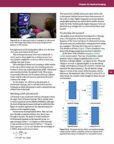
Figure 32.21 An expectant mother undergoes an ultrasound scan. The image of her baby is built up by computer and appears on the monitor.
first appearance in the family photo album is in the form of an ante-natal (before birth) scan!
This technique has many other uses in medicine. It can be used to detect gallstones or kidney stones (two very painful complaints), so men as well as women may undergo this type of scan.
The technique of ultrasound scanning is rather similar to the way in which sailors use echo sounding and echo location to detect the seabed and shoals of fish. Ultrasound waves are directed into the patient’s body. These waves
are partially reflected at the boundaries between different tissues and the reflected waves are detected and used to construct the image.
In this chapter, we will look at the principles of ultrasound scanning, and we will also look at another technique in which ultrasound is used to measure the rate of blood flow in the body.
Working with ultrasound
Ultrasound is any sound wave that has a frequency above the upper limit of human hearing. This is usually taken to mean frequencies above 20 kHz (20 000 Hz), although the limit of hearing decreases with age to well below this figure. In medical applications, the typical frequencies used are in the megahertz range.
Sound waves are longitudinal waves. They can only pass through a material medium; they cannot pass through a vacuum. The speed of sound (and hence
of ultrasound) depends on the material. In air, it is approximately 330 m s−1; it is higher in solid materials.
A typical value for body tissue is 1500 m s−1. Using the wave equation v = f λ, we can calculate the wavelength of 2.0 MHz ultrasound waves in tissue:
λ = v = 1500 ×106 f 2.0
This means that 2.0 MHz ultrasound waves will be able
to distinguish detailed features whose dimensions are of the order of 1 mm. Higher-frequency waves have shorter wavelengths and these are used to detect smaller features inside the body. Unfortunately, higher frequency waves are absorbed more strongly and so a more intense beam must be used.
Producing ultrasound
Like audible sound, ultrasound is produced by a vibrating source. The frequency of the source is the same as the frequency of the waves it produces. In ultrasound scanning, ultrasonic waves are produced by a varying electrical voltage in a transducer. The same device also acts as a detector. (You should recall from Chapter 25 that a transducer is any device that changes energy from one form to another.)
At the heart of the transducer is a piezo-electric crystal, such as quartz. This type of crystal has a useful property: when a voltage is applied across it in one direction, it shrinks slightly – see Figure 32.22a. When the voltage is reversed, it expands slightly. So an alternating voltage with frequency f causes the crystal to contract and expand at the same frequency f. We say that the voltage induces a strain in the crystal. In the best piezo-electric substances, the maximum value of strain is about 0.1%; in other words, the crystal’s width changes by about one part in a thousand.
a
= 7.5 × 10⁻⁴ m ≈ 1 mm
b
+–
applied voltage
applied stress
induced strain
+– +– +– +– +– +–
induced e.m.f.
Figure 32.22 The piezo-electric effect. a An applied voltage causes a piezo-electric crystal to contract or expand. b An applied stress causes an induced e.m.f. across the crystal.
517
