
Page 527 - Physics Coursebook 2015 (A level)
P. 527
Chapter 32: Medical imaging
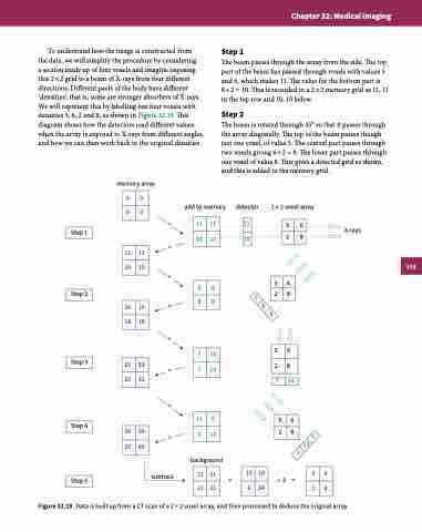
To understand how the image is constructed from
the data, we will simplify the procedure by considering
a section made up of four voxels and imagine exposing this 2 × 2 grid to a beam of X-rays from four different directions. Different parts of the body have different ‘densities’; that is, some are stronger absorbers of X-rays. We will represent this by labelling our four voxels with densities 5, 6, 2 and 8, as shown in Figure 32.19. This diagram shows how the detectors read different values when the array is exposed to X-rays from different angles, and how we can then work back to the original densities.
memory array
+ =
Step 1
The beam passes through the array from the side. The top part of the beam has passed through voxels with values 5 and 6, which makes 11. The value for the bottom part is 8+2 = 10. This is recorded in a 2×2 memory grid as 11, 11 in the top row and 10, 10 below.
Step 2
The beam is rotated through 45° so that it passes through the array diagonally. The top of the beam passes though just one voxel, of value 5. The central part passes through two voxels giving 6 + 2 = 8. The lower part passes through one voxel of value 8. This gives a detected grid as shown, and this is added to the memory grid.
0
0
0
0
add to memory
detector
2 × 2 voxel array
11
56
X-rays
Step 1
10
28
11
11
10
10
+ =8
+ =
+
=
56
28
5
8
8
8
Step 2
Step 3
5
8
16
19
18
18
5
6
7
14
7
14
23
33
25
32
2
7 14
8
13
6
2
13
56
28
Step 4
36
39
27
45
background subtract
6 13
2
21
21
21
21
15
18
6
24
5
6
2
8
= ÷3=
Figure 32.19 Data is built up from a CT scan of a 2 × 2 voxel array, and then processed to deduce the original array.
Step 5
11
10
11
10
515
