
Page 12 - HBC 2017 - Final
P. 12
118 International Orthopaedics (SICOT) (2008) 32:115–119
Table 2 Fusion criteria used for 136 patients who underwent PLF [4]. No matter what the aetiology of the LS, patients usually
with laminectomy bone chips present with a persistent dull low-back pain with radicul-
Criteria opathy, which increases with activity and decreases with
rest, low-back stiffness, tight hamstrings and intermittent
Solid fusion Failed fusion (non-union) claudication. The mainstay of treatment is conservative,
with rest, use of NSAIDs, physical therapy and the wearing
Bridging intertransverse bone Absence of bridging
of a body brace. Surgical intervention is only performed
interteranverse bone
No motion on lat flexion- Presence of motion on lat flexion- when there is failure of conservative treatment for at least
extension radiographs extension radiographs one year. Surgical treatment of LS may be done via
Subsidence to <75% of Subsidence to >75% of original either an anterior or posterior approach. After the introduc-
original disc space height disc space height tion of instrumentation for spinal reduction in the 1960s,
operative management for LS is commonly performed via a
posterior decompression laminectomy with posterolateral
forwardinrelationshiptothe vertebra below[1, 4].
fusion and reduction of the slipped vertebra with spinal
However, with the aging population found in an industrial
instrumentation.
country like Taiwan, the prevalence of degenerative
Fusion is the most important factor in the successful
spondylolisthesis has grown. The pathology of degenerative
treatment of LS, with autogenous, allograft, dimineralised
spondylolisthesis is different from that of isthmic spondy-
bone matrix (DBM) and other graft extenders, such as
lolisthesis; that is, the pars in degenerative spondylolis-
calcium phosphate, as options to achieve this objective.
thesis remains intact, with the forward slippage caused by
Overall, present studies show that an autogenous graft
arthritic changes in the zygapophyseal joints between two
provides the best fusion, because of its osteogenic,
vertebrae associated with degeneration of the disc at that
osteoconductive, and osteoinductive properties. An allo-
level [9–11]. The most frequent site of pathology is
graft bone, which has low or no osteogenicity and weak
between L4 and L5, with L3 next in order of occurrence
osteoinductive properties, is very poor in stimulating
fusion. It is also reported that an allograft bone has
increased immunogenicity, increasing its risk for disease
transmission and resorbs more rapidly than an autogenous
graft. The DBM and graft extenders contain proteins that
stimulate bone formation and have successfully fused
spines in animal studies, but at present there is no
sufficient information to prove that they effectively
stimulate successfull fusion in the human spine. They
are also expensive, and are not recommended for use
without addition of the patient’s own bone. So far, the
most popular donor site for autogenous graft is the iliac
crest. In our study, we discovered that laminectomy bone
chips are excellent for PLF, both in quantity and quality.
In the case of iliac crest bone harvest, the donor site is at
risk of complications, such as large haematoma, wound
infection, disabling donor wound pain, unsightly scars,
meralgia paraesthesia, pelvic fracture (high in patients
with osteoporosis), herniation at the harvest site, suture
rejection with prolonged sterile drainage and seroma. All
of these can lengthen hospital stay and may require
additional surgery, leading to additional cost of treatment
[2]. However, these co-morbidities were found to be
extremely variable by different authors. As for the
technique we have describe, our patients did not encounter
any of the above mentioned co-morbidities with a short
operative time and minimal blood loss noted.
In our series, we achieved a fusion rate of 94.85% (129/
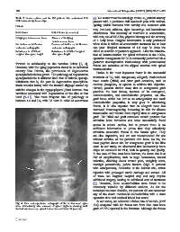
Fig. 4 Plain anteroposterior radiograph of the lumbo-sacral spine
taken immediately after removal of spinal implants showing well- 136) compared with the fusion rate from iliac crest bone
formed intertransverse spinal fusion mass at L4 to L5 (arrows) graft of 97% reported in the literature. Although the fusion