
Page 46 - Power of Stem Cells- arthritis and regeneration
P. 46
Luque-Campos et al. MSCs and Memory T Cells in RA
organism against extracellular pathogens, including Gram- Treg correlated with the DAS28 (127). However, despite the
negative bacteria, mycobacteria, and fungi (109). However, increased number of Treg in the synovial fluid, inflammation
their deregulation is associated with the generation of auto- is maintained suggesting an alteration of their functions in
immune diseases including RA (109). On the other side, it RA patients. This was confirmed by a body of studies that
is well known that human Treg cells play a central role in has demonstrated by the reduced regulatory functions of Treg
the maintenance of immune homeostasis and immunological derived from the peripheral blood (128–131) and the synovial
self-tolerance (110). Treg cells exert potent immunosuppressive fluid of RA patients (132). In line with these studies, Treg
effects over effector T-cell proliferation and cytokine production cells isolated from patients with active RA did not inhibit the
through cytokine-independent mechanism requiring cell-to- secretion of pro-inflammatory cytokine such as IFN-γ and TNFα
cell contact. Treg cells are characterized by high expression released by T effector cells (127–130, 133). Notoriously, TNFα
level of CD25 (also referred as CD25 bright cells) and more can inhibit the suppressive function of Treg (129) suggesting that
specifically, intracellular expression of the transcription factor RA synovial fluid enriched in pro-inflammatory convert memory
FoxP3 (111, 112). Moreover, Treg are characterized by a low Treg cells into cells producing pro-inflammatory cytokines such
expression of CD127 (IL-7 receptor alpha-chain) (113), and a as IL-17 unable to exert regulatory functions (134). An increased
−
down-regulation of CD127 which is associated with regulatory percentage of memory CD45RA Foxp3 low non-regulatory T
function acquisition (114). The imbalance between Th17 and cells was reported in RA synovial fluid while it did not change
Treg cells has been largely associated with the RA pathogenesis in the peripheral blood of patients (55). Memory non-Treg
due to their close differentiation pathways but their completely cells produce IL-2, IFN-γ, and IL-17 and express high levels of
opposite function. (115, 116). Indeed, Th17 cells are implicated RORC (135, 136).
+
in RA development and progression and high levels of IL-17 MSCs are potent inhibitors of CD4 T-bet CD183 +
+
+
have been reported in the synovial fluid of RA patients which is (Th1) and CD4 RORγt CD161 + (Th17) cells proliferation
+
positively correlated with the severity of the disease (117–120). and significantly reduce their capacity to produce pro-
Furthermore, IL-17 is mainly produced by CD4 CD45RO + inflammatory cytokines such as IFN-γ, TNFα, and IL-1β
+
memory T cell (121, 122). Another molecule, the chemokine (Th1) and IL-17A and IL-22 (Th17) (80). Indeed, using memory
+
+
receptor CCR6, is expressed by memory Th17 cells and associated CD4 CD45RO CCR6 + positive cells (Th17 cells), human
with their capacity to migrate toward inflammatory joints in BM-MSCs have been shown to induce the generation of Th17
response to CCL20 highly produced by T cells and synoviocytes cells with regulatory features in an inflammatory environment
+
(123, 124). On the other hand, CD4 CD25 high Treg cells are characterized by a decrease in RORC expression, an increase of
predominantly memory cells in the synovial fluid which is FoxP3 expression and the acquisition of immunosuppressive
◦w
enriched with CD4 CD25 CD127l FoxP3 + Treg cells in the functions (137).
+
+
synovial fluid of RA patients (111, 125, 126). Furthermore, Likewise, various studies have shown that MSCs have the
while the percentage of memory Treg cells subsets significantly capacity to increase the percentage of Treg cells in vitro in co-
increased in the synovial fluid of RA patients, it did not change in culture in mixed lymphocyte reactions (MLR) (138, 139). MSCs-
their peripheral blood, and this increased frequency of memory derived PGE2 and transforming growth factor beta 1 (TGFβ1)
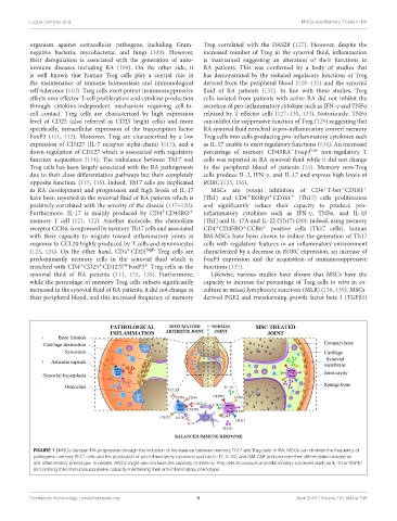
FIGURE 1 | MSCs dampen RA progression through the induction of the balance between memory Th17 and Treg cells. In RA, MSCs can diminish the frequency of
pathogenic memory Th17 cells and the production of pro-inflammatory cytokines such as IL-17, IL-22, and GM-CSF and promote their differentiation toward an
anti-inflammatory phenotype. In parallel, MSCs might also increase the capacity of memory Treg cells to produce anti-inflammatory cytokines such as IL-10 or TGFβ1
and prolong their immunosuppressive capacity maintaining their anti-inflammatory phenotype.
Frontiers in Immunology | www.frontiersin.org 6 April 2019 | Volume 10 | Article 798