
Page 12 - Gastric pentadecapeptide BPC 157
P. 12
118
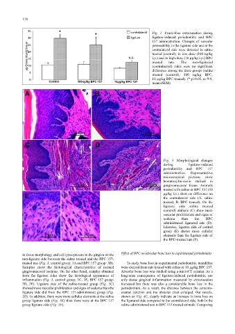
35
* contralateral Fig. 2. Evans-blue extravasation during
30 * ligature ligature-induced periodontitis and BPC
157 administration. Changes of vascular
µg Evans blue/g tissue 20 N.S. contralateral side were detected in saline
permeability in the ligature side and at the
25
treated (control), in low dose (100 ng/kg
i.p.) and in high-dose (10 µg/kg i.p.) BPC
15
treated
rats.
The
non-ligatured
10
difference among the three groups (saline
5 (contralateral) sides were no significant
treated (control), 100 ng/kg BPC,
0 10 µg/kg BPC treated). (* p<0.05, n=9-9,
Control 100ng/kg BPC 157 10µg/kg BPC 157
mean±SEM)
Fig. 3. Morphological changes
during ligature-induced
periodontitis and BPC 157
administration. Representative
microscopical pictures show
hematoxylin-eosin stained in
gingivomucosal tissue. Animals
treated with saline or BPC 157 (10
µg/kg i.p.) show no difference on
the contralateral side (A: saline
treated; B: BPC treated). On the
ligature side saline treated
(control) animals (C) show more
vascular proliferation and signs of
oedema than the BPC
administrated ligatured rats (D).
Likewise, ligature side of control
group (E) shows more cellular
elements than the ligature side of
the BPC-treated rats (F).
Effect of BPC on alveolar bone loss in experimental periodontitis
in tissue morphology and cell types present in the gingiva on the
non-ligature side between the saline treated and the BPC 157-
treated rats (Fig. 3, control group: 3A and BPC 157 group: 3B). To study bone loss in experimental periodontitis, mandibles
Samples show the histological characteristics of normal were excised from rats treated with saline or 10 µg/kg BPC 157.
gingivomucosal sections. On the other hand, samples obtained Alveolar bone loss was studied using a microCT scanner. As a
from the ligature sides show the histological appearance of long-term consequence of ligature-induced periodontitis, not
inflammation (Fig. 3, control group: 3C, 3E, BPC 157 group: only tissue gingival inflammation measured by extravasation
3D, 3F). Ligature side of the saline-treated group (Fig. 3C) increased but there was also a considerable bone loss in the
showed more vascular proliferation and signs of oedema than the periodontium. As a result, the distance between the cemento-
ligature side did from the BPC 157-administered group (Fig. enamel junction and the alveolar crest enlarged. Our results,
3D). In addition, there were more cellular elements at the saline shown on Fig. 4C, clearly indicate an increase in bone loss on
group ligature side (Fig. 3E) than there were at the BPC 157 the ligatured side compared to the contralateral side, both in the
group ligature side (Fig. 3F). saline-administered and in BPC 157-treated animals. Comparing