
Page 21 - Atlas of Small Animal CT and MRI
P. 21
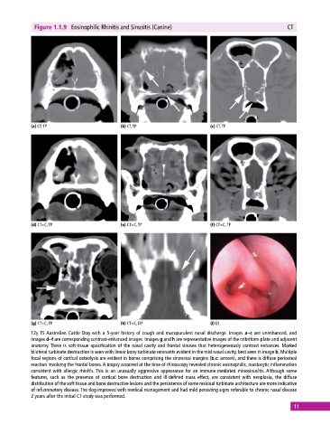
Figure 1.1.9  Eosinophilic Rhinitis and Sinusitis (Canine)                                  CT


            (a) CT, TP                       (b) CT, TP                       (c) CT, TP


            (d) CT+C, TP                     (e) CT+C, TP                     (f) CT+C, TP


            (g) CT+C, TP                     (h) CT+C, DP                     (i) ES
            12y FS Australian Cattle Dog with a 5‐year history of cough and mucopurulent nasal discharge. Images a–c are unenhanced, and
            images d–f are corresponding contrast‐enhanced images. Images g and h are representative images of the cribriform plate and adjacent
            anatomy. There is soft‐tissue opacification of the nasal cavity and frontal sinuses that heterogeneously contrast enhances. Marked
              bilateral turbinate destruction is seen with linear bony turbinate remnants evident in the mid‐nasal cavity, best seen in image b. Multiple
            focal regions of cortical osteolysis are evident in bones comprising the sinonasal margins (b,c: arrows), and there is diffuse periosteal
            reaction involving the frontal bones. A biopsy acquired at the time of rhinoscopy revealed chronic eosinophilic, mastocytic inflammation
            consistent with allergic rhinitis. This is an unusually aggressive appearance for an immune‐mediated rhinosinusitis. Although some
              features, such as the presence of cortical bone destruction  and ill‐defined mass  effect, are consistent with neoplasia, the diffuse
              distribution of the soft tissue and bone destructive lesions and the persistence of some residual turbinate architecture are more indicative
            of inflammatory disease. The dog improved with medical management and had mild persisting signs referable to chronic nasal disease
            2 years after the initial CT study was performed.
                                                                                                             11