
Page 246 - Atlas of Small Animal CT and MRI
P. 246
236 Atlas of Small Animal CT and MRI
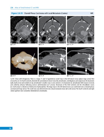
Figure 2.8.15 Choroid Plexus Carcinoma with Local Metastasis (Canine) MR
(a) T2, TP (b) T1, TP (c) T1+C, TP
(d) GP, TP (e) T1, SP (f) T1+C, SP
7y MC Chow with tetraparesis. There is a large, T1 and T2 hyperintense ovoid mass in the left lateral recess (a,b,e: large arrow) that
encroaches on the fourth ventricle, distends the contralateral lateral recess (b: arrowheads), and displaces and compresses the brainstem
(a,b: small arrow) and cerebellum (a: arrowhead). A smaller mass is seen ventral to the brainstem (e: arrowhead). Both masses intensely
and uniformly enhance following contrast administration (c,f: arrows). Distension of the third ventricle and the infundibular recess
(e: small arrow) is indicative of obstructive hydrocephalus. The large mass in the left lateral recess was confirmed to be a choroid plexus
carcinoma (d: large arrow). The small mass was determined to be a local metastatic lesion (d: small arrow). The fourth ventricle and right
lateral aperture were somewhat distended (d: arrowheads).
236