
Page 31 - Atlas of Small Animal CT and MRI
P. 31
Nasal Cavity and Paranasal Sinuses 21
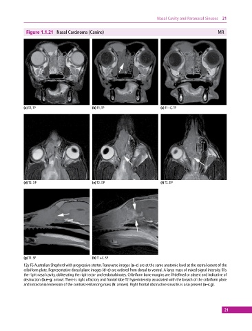
Figure 1.1.21 Nasal Carcinoma (Canine) MR
(a) T2, TP (b) T1, TP (c) T1+C, TP
(d) T2, DP (e) T2, DP (f) T2, DP
(g) T1, SP (h) T1+C, SP
12y FS Australian Shepherd with progressive stertor. Transverse images (a–c) are at the same anatomic level at the rostral extent of the
cribriform plate. Representative dorsal plane images (d–e) are ordered from dorsal to ventral. A large mass of mixed‐signal intensity fills
the right nasal cavity, obliterating the right ecto‐ and endoturbinates. Cribriform bone margins are ill‐defined or absent and indicative of
destruction (b,e–g: arrow). There is right olfactory and frontal lobe T2 hyperintensity associated with the breach of the cribriform plate
and intracranial extension of the contrast‐enhancing mass (h: arrows). Right frontal obstructive sinusitis is also present (a–c,g).
21