
Page 33 - Atlas of Small Animal CT and MRI
P. 33
Nasal Cavity and Paranasal Sinuses 23
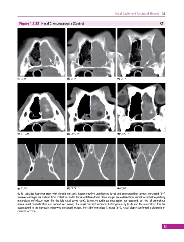
Figure 1.1.23 Nasal Chondrosarcoma (Canine) CT
(a) CT, TP (b) CT, TP (c) CT, TP
(d) CT+C, TP (e) CT+C, TP (f) CT+C, TP
(g) CT, DP (h) CT, DP (i) CT, DP
6y FS Labrador Retriever cross with chronic epistaxis. Representative unenhanced (a–c) and corresponding contrast‐enhanced (d–f)
transverse images are ordered from rostral to caudal. Representative dorsal plane images are ordered from dorsal to ventral. A partially
mineralized soft‐tissue mass fills the left nasal cavity (a–c). Extensive turbinate destruction has occurred, but foci of amorphous
intralesional mineralization are evident (a,c: arrow). The mass contrast enhances heterogeneously (d–f), and the mineralized foci are
accentuated in the narrowly windowed enhanced images. The cribriform plate is intact (g–i). Nasal biopsy confirmed a diagnosis of
chondrosarcoma.
23