
Page 417 - Atlas of Small Animal CT and MRI
P. 417
Pleural Space 407
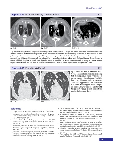
Figure 4.2.11 Metastatic Mammary Carcinoma (Feline) CT
(a) CT, TP (b) CT+C, TP (c) CT+C, TP
12y FS Domestic Longhair with progressive respiratory distress. Representative CT images include an unenhanced (a) and corresponding
contrast‐enhanced (b) transverse image of the cranial thorax and an additional transverse image at the level of the midthorax (c). The
patient is positioned in dorsal recumbency, and images are displayed in that orientation. A heterogeneous contrast‐enhancing mass is
present in the right ventral thoracic wall and extends into the ventral mediastinum (a,b: arrow). Moderate bilateral pleural effusion is
present with fluid distributed primarily in the dependent thorax (c: asterisks). The ventral lung is atelectatic (c: arrow) with nondependent
regions better aerated. The mass was confirmed to be a subpleural metastatic mammary carcinoma with pleural effusion.
Figure 4.2.12 Pleural Fibrosis (Canine) CT
8y FS Shiba Inu with a retrobulbar mass.
CT was performed as a metastasis‐screening
test. Heterogeneous pleural thickening is
present involving visceral pleura of multiple
lung lobes bilaterally (a,b: arrowheads).
There is no suggestion of a pleural effusion
component, which suggests pleural changes
are inactive. Pleural thickening was thought
to represent residual pleural fibrosis from
previous inflammatory disease.
(a) CT, TP (b) CT, TP
References 5. Lee N, Won S, Choi M, Kim J, Yi K, Chang D, et al. CT thoracic
duct lymphography in cats by popliteal lymph node iohexol injec-
1. Au JJ, Weisman DL, Stefanacci JD, Palmisano MP. Use of computed tion. Vet Radiol Ultrasound. 2012;53: 174–180.
tomography for evaluation of lung lesions associated with sponta- 6. Swinbourne F, Baines EA, Baines SJ, Halfacree ZJ. Computed
neous pneumothorax in dogs: 12 cases (1999–2002). J Am Vet Med tomographic findings in canine pyothorax and correlation with
Assoc. 2006;228: 733–737. findings at exploratory thoracotomy. J Small Anim Pract. 2011;52:
2. Lipscomb V, Brockman D, Gregory S, Baines S, Lamb CR. CT 203–208.
scanning of dogs with spontaneous pneumothorax. Vet Rec. 7. Schultz RM, Zwingenberger A. Radiographic, computed tomographic,
2004;154: 344. and ultrasonographic findings with migrating intrathoracic grass
3. Dechman G, Mishima M, Bates JH. Assessment of acute pleural awns in dogs and cats. Vet Radiol Ultrasound. 2008;49: 249–255.
effusion in dogs by computed tomography. J Appl Physiol. 1994;76: 8. Echandi RL, Morandi F, Newman SJ, Holford A. Imaging diagnosis –
1993–1998. canine thoracic mesothelioma. Vet Radiol Ultrasound. 2007;48:
4. Johnson EG, Wisner ER, Kyles A, Koehler C, Marks SL. Computed 243–245.
tomographic lymphography of the thoracic duct by mesenteric 9. Reetz JA, Buza EL, Krick EL. CT features of pleural masses and
lymph node injection. Vet Surg. 2009;38: 361–367. nodules. Vet Radiol Ultrasound. 2012;53: 121–127.
406 407