
Page 421 - Atlas of Small Animal CT and MRI
P. 421
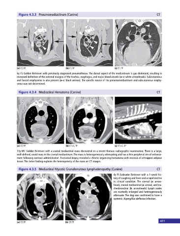
Figure 4.3.3 Pneumomediastinum (Canine) CT
(a) CT, TP (b) CT, TP (c) CT, TP
6y FS Golden Retriever with previously diagnosed pneumothorax. The dorsal aspect of the mediastinum is gas distended, resulting in
increased definition of the external margins of the trachea, esophagus, and major blood vessels (a–c: white arrowheads). Subcutaneous
and fascial emphysema is also present (a–c: black arrows). The specific source of the pneumomediastinum and subcutaneous emphy
sema was not determined.
Figure 4.3.4 Mediastinal Hematoma (Canine) CT
(a) CT, TP (b) CT+C, TP (c) CT+C, SP
10y MC Golden Retriever with a cranial mediastinal mass discovered on a recent thoracic radiographic examination. There is a large,
well‐defined, ovoid mass in the cranial mediastinum. The mass is heterogeneously attenuating and has a thin peripheral rim of enhance
ment following contrast administration. Excisional biopsy revealed a chronic organizing hematoma with necrosis of entrapped adipose
tissue. This latter finding explains the heterogeneity of the mass on CT images.
Figure 4.3.5 Mediastinal Mycotic Granulomatous Lymphadenopathy (Canine) CT
4y FS Labrador Retriever with a 1‐week his
tory of coughing and fever and a rapid decline
in clinical condition. The sternal (a: arrow
head), cranial mediastinal (a: arrow), and tra
cheobronchial (b: arrowheads) lymph nodes
are markedly enlarged and heterogeneously
attenuate. The dog was confirmed to have a
systemic Aspergillus deflectus infection.
411
(a) CT, TP (b) CT, TP