
Page 423 - Atlas of Small Animal CT and MRI
P. 423
Mediastinum and esophagus 413
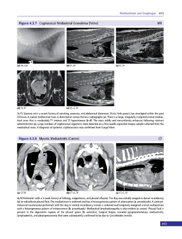
Figure 4.3.7 Cryptococcal Mediastinal Granuloma (Feline) MR
(a) DX, LAT (b) T1, SP (c) T2, SP
(d) T2, TP (e) T1+C, SP
3y FS Siamese with a recent history of vomiting, anorexia, and abdominal distension. Pelvic limb paresis has developed within the past
24 hours. A cranial mediastinal mass is detected on survey thoracic radiographs (a). There is a large, irregularly margined cranial medias
tinal mass that is moderately T1 intense and T2 hyperintense (b–d). The mass mildly and nonuniformly enhances following contrast
administration (e). Large numbers of cryptococcal organisms were detected on a fine‐needle aspiration biopsy sample collected from the
mediastinal mass. A diagnosis of systemic cryptococcosis was confirmed from fungal titers.
Figure 4.3.8 Mycotic Mediastinitis (Canine) CT
(a) CT, TP (b) CT+C, TP (c) CT+C, SP
4y M Rottweiler with a 3‐week history of lethargy, inappetence, and pleural effusion. The dog was initially imaged in dorsal recumbency
(a) to redistribute pleural fluid. The mediastinum is widened and has a heterogeneous pattern of attenuation (a: arrowheads). A contrast‐
enhanced examination performed with the dog in sternal recumbency reveals a widened and irregularly margined cranial mediastinum
with a heterogeneous pattern of enhancement (b: arrowheads). Mediastinal lymphadenopathy is also evident (c: arrow). Pleural fluid is
present in the dependent regions of the pleural space (b: asterisks). Surgical biopsy revealed pyogranulomatous mediastinitis,
lymphadenitis, and pleuropneumonia that were subsequently confirmed to be due to Coccidioides immitis.
413