Page 136 - YORAM RUDY BOOK FINAL
P. 136
P. 136
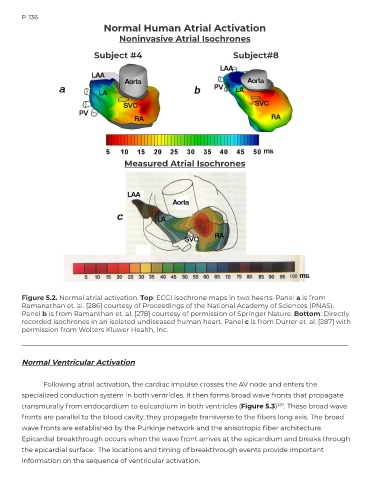
                                   Normal Human Atrial Activation

                                        Noninvasive Atrial Isochrones

                                 Subject #4                                      Subject#8


                                          Measured Atrial Isochrones


        Figure 5.2. Normal atrial activation. Top: ECGI isochrone maps in two hearts. Panel a is from
        Ramanathan et. al. [286] courtesy of Proceedings of the National Academy of Sciences (PNAS).
        Panel b is from Ramanthan et. al. [278] courtesy of permission of Springer Nature. Bottom: Directly
        recorded isochrones in an isolated undiseased human heart. Panel c is from Durrer et. al. [287] with
        permission from Wolters Kluwer Health, Inc.


        Normal Ventricular Activation


               Following atrial activation, the cardiac impulse crosses the AV node and enters the
        specialized conduction system in both ventricles. It then forms broad wave fronts that propagate

        transmurally from endocardium to epicardium in both ventricles (Figure 5.3)          287 . These broad wave
        fronts are parallel to the blood cavity; they propagate transverse to the fibers long axis. The broad
        wave fronts are established by the Purkinje network and the anisotropic fiber architecture.

        Epicardial breakthrough occurs when the wave front arrives at the epicardium and breaks through
        the epicardial surface.  The locations and timing of breakthrough events provide important
        information on the sequence of ventricular activation.

