
Page 47 - Clinical Manual of Small Animal Endosurgery
P. 47
Diagnostic Arthroscopy 35
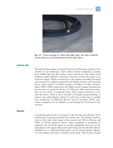
Fig. 2.3 Close-up image of a fibre-optic light cable. The cable should be
wound gently to avoid breaking the delicate glass fibres.
Camera unit
The arthroscopic image is projected from the arthroscope eyepiece to the
monitor via an endoscopic video camera system comprising a camera
head, which clips onto the eyepiece, and a control unit. The camera head
contains a light-responsive electronic chip that converts the image to an
electronic signal, which is transferred to the camera controller, processed
and exported as a signal recognised by the display monitor and recording
device. Most camera controllers produce a number of export formats.
Super VHS (s-VHS) output gives the highest-quality image reproduction
but attention is required in the use of s-VHS-type cable connections since
they are not robust. A composite video cable output is therefore a suit-
able alternative which is more resistant to damage during handling. A
camera unit with multiple outputs of each type is ideal since it enables
easy connection of additional devices such as recorders (DVD, cam-
corder, computer, etc.) in addition to maintaining the connection to the
monitor.
Monitor
A medical-grade monitor is necessary to get the best reproduction of the
arthroscopic image generated by the camera unit. The monitor needs to
work on the same video signal as the camera unit (PAL in Europe and
NTSC in North America); hence, when equipment is purchased or
replaced, care should be taken to purchase equipment that runs on the
appropriate video signal. Flat-screen technology has become the industry
standard but a traditional high-quality colour medical-grade cathode-
ray-tube monitor provides a perfectly good image. The monitor should