
Page 117 - Rapid Review of ECG Interpretation in Small Animal Practice, 2nd Edition
P. 117
Answer 47 New General-Level ECG Cases
Answer 47
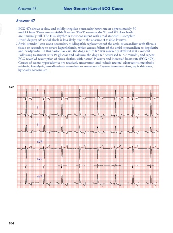
VetBooks.ir 1 ECG 47a shows a slow and mildly irregular ventricular heart rate at approximately 30
and 35 bpm. There are no visible P waves. The T waves in the V1 and V3 chest leads
are unusually tall. The ECG rhythm is most consistent with atrial standstill. Complete
(third-degree) AV nodal block is less likely due to the absence of visible P waves.
2 Atrial standstill can occur secondary to idiopathic replacement of the atrial myocardium with fibrous
tissue or secondary to severe hyperkalemia, which causes failure of the atrial myocardium to depolarize
+
and bradycardia. In this particular case, the dog’s serum K was markedly elevated at 8.7 mmol/L.
+
Following treatment with IV glucose and calcium, the dog’s K decreased to 7.7 mmol/L, and repeat
ECG revealed resumption of sinus rhythm with normal P waves and increased heart rate (ECG 47b).
Causes of severe hyperkalemia are relatively uncommon and include ureteral obstruction, metabolic
acidosis, hemolysis, complications secondary to treatment of hyperadrenocorticism, or, in this case,
hypoadrenocorticism.
47b
I
II
III
aVR
aVL
aVF
104