
Page 121 - Rapid Review of ECG Interpretation in Small Animal Practice, 2nd Edition
P. 121
Answer 49 New General-Level ECG Cases
Answer 49
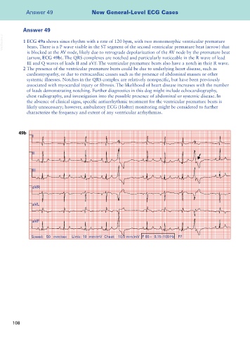
VetBooks.ir 1 ECG 49a shows sinus rhythm with a rate of 120 bpm, with two monomorphic ventricular premature
beats. There is a P wave visible in the ST segment of the second ventricular premature beat (arrow) that
is blocked at the AV node, likely due to retrograde depolarization of the AV node by the premature beat
(arrow, ECG 49b). The QRS complexes are notched and particularly noticeable in the R wave of lead
III and Q waves of leads II and aVF. The ventricular premature beats also have a notch in their R wave.
2 The presence of the ventricular premature beats could be due to underlying heart disease, such as
cardiomyopathy, or due to extracardiac causes such as the presence of abdominal masses or other
systemic illnesses. Notches in the QRS complex are relatively nonspecific, but have been previously
associated with myocardial injury or fibrosis. The likelihood of heart disease increases with the number
of leads demonstrating notching. Further diagnostics in this dog might include echocardiography,
chest radiography, and investigation into the possible presence of abdominal or systemic disease. In
the absence of clinical signs, specific antiarrhythmic treatment for the ventricular premature beats is
likely unnecessary; however, ambulatory ECG (Holter) monitoring might be considered to further
characterize the frequency and extent of any ventricular arrhythmias.
49b
I
II
III
aVR
aVL
aVF
Speed: 50 mm/sec Limb: 10 mm/mV Chest: 10.0 mm/mV F 60~ 0.15–100 Hz P?
108