
Page 223 - Fluid, Electrolyte, and Acid-Base Disorders in Small Animal Practice
P. 223
Disorders of Magnesium: Magnesium Deficit and Excess 213
complexed form (15% to 25%). Unlike calcium, which is transepithelial concentration gradient generally favors
approximately 40% protein bound, magnesium is only absorption of magnesium from the gut and is influenced
20% to 30% bound to protein and so is less affected by by the gut intraluminal ionized magnesium concentra-
changes in albumin concentration. 90,124 Inside the cell, tion (chelated or complexed magnesium species do not
magnesium is complexed to many organic compounds contribute). Thus, total dietary intake of magnesium
whereitplaysapivotalrole.Currentestimatesindicatethat and intake of dietary constituents that influence the
only about 1% to 2% of the intracellular magnesium is amount of magnesium that is complexed or chelated
present in the ionized or free form. Presumably, may influence the net absorption of magnesium. The
magnesium also shifts between the free and complexed transepithelial voltage gradient is created by net move-
intracellular forms as well, but the precise regulatory ment of salt and water. A small positive intraluminal volt-
mechanisms governing those shifts are not understood age results in a small force favoring transepithelial cation
at this time. movement. Solvent drag created by sodium and water
reabsorption will also result in transepithelial movement
GASTROINTESTINAL HANDLING OF of magnesium and other ions. Water and salt reabsorption
MAGNESIUM from the gut therefore has a significant influence on mag-
The primary site of magnesium absorption appears to be nesium absorption.
the ileum, but the jejunum and colon also contribute sub- The permeability of the paracellular tight junctions is
stantially to net absorption. 70,71 The mechanisms of mag- currently an area of intense study and interest. Numerous
nesium absorption from the ileum are the most well proteins exist in the tight junction that serve as ion
studied at this time. Much research remains yet to channels and influence permeability of many ions. Spe-
completely understand the complexities of gastrointesti- cific magnesium ion channels in the gut epithelial tight
nal magnesium absorption. Several key mechanisms are junctions have not been conclusively identified. Proteins
currently well understood. Two pathways for intestinal regulating magnesium movement through the renal epi-
magnesium absorption exist: an unsaturable passive thelial tight junctions have been identified (paracellin-1
paracellular route and a saturable active transcellular [PCLN-1]), leading to speculation that a similar protein
route (Fig. 8-1). 70,71,80 The paracellular movement of may exist in the gut as well. Once identified conclusively,
magnesium occurs through the tight junctions between further study will be required to determine if this tight
epithelial cells. The driving forces for paracellular magne- junction protein is selectively permeable and under what
sium movement are: transepithelial magnesium concen- influences such selectivity is expressed.
tration gradient, the transepithelial voltage gradient Active transcellular magnesium movement from the
formed by salt and water absorption, and the permeability gut is an area of very recent and exciting discovery. Study
of the tight junctions to magnesium. 81 The of numerous inherited conditions of impaired magnesium
handling in humans led to an improved understanding of
magnesium transport across the gut epithelium and a
hypothesisthatseveralmagnesiumtransportproteinsexist
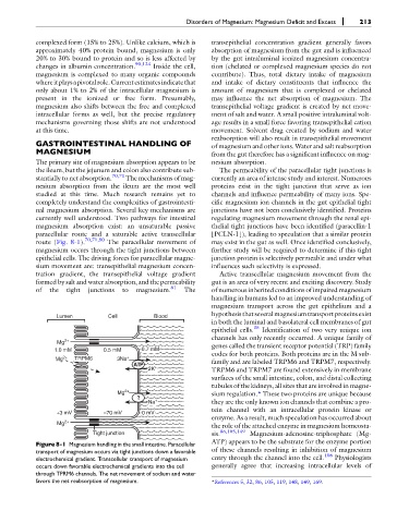
Lumen Cell Blood
in both the luminal and basolateral cell membranes of gut
epithelial cells. 35 Identification of two very unique ion
channels has only recently occurred. A unique family of
Mg 2+
1.0 mM 0.5 mM 0.7 mM genes called the transient receptor potential (TRP) family
codes for both proteins. Both proteins are in the M sub-
Mg 2+ TRPM6 3Na +
ATP family and are labeled TRPM6 and TRPM7, respectively.
2K + TRPM6 and TRPM7 are found extensively in membrane
surfaces of the small intestine, colon, and distal collecting
tubules of the kidneys, all sites that are involved in magne-
Mg 2+ sium regulation.* These two proteins are unique because
?
Na + they are the only known ion channels that combine a pro-
tein channel with an intracellular protein kinase or
+3 mV –70 mV 0 mV
enzyme. As a result, much speculation has occurred about
Mg 2+
the role of the attached enzyme in magnesium homeosta-
Tight junction sis. 86,105,149 Magnesium-adenosine-triphosphate (Mg-
Figure 8-1 Magnesium handling in the small intestine. Paracellular ATP) appears to be the substrate for the enzyme portion
transport of magnesium occurs via tight junctions down a favorable of these channels resulting in inhibition of magnesium
electrochemical gradient. Transcellular transport of magnesium entry through the channel into the cell. 106 Physiologists
occurs down favorable electrochemical gradients into the cell generally agree that increasing intracellular levels of
through TPRM6 channels. The net movement of sodium and water
favors the net reabsorption of magnesium. *References 5, 32, 86, 105, 119, 148, 149, 169.