Page 249 - Fluid, Electrolyte, and Acid-Base Disorders in Small Animal Practice
P. 249
240        ACID-BASE DISORDERS


                           [H ] +  120 100  80 70
                           pH  6.92 7.00   60
               100                  7.10 7.15 7.22                  34
                                               50
                                                     +
                                             7.30
                                                                    32
                                                   40
                                                    [H ] nEq/L
                                   Acute resp. acidosis  Chronic resp. acidosis  7.52  HCO 3  mEq/L  28
               80                                7.40   30          30          Whole blood
             PaCO 2  mm Hg  40        Metabolic  alkalosis  7.70  20  26                       Whole body
               60

                                                                    24
                         Chronic resp. alkalosis
                      Metabolic acidosis Acute resp. alkalosis  8.00  10  22
               20                                                   20
                                                                           40     60    80    100    120   140
                                                +    PaCO 2
                                               [H ] = 24                              pCO 2  (mm Hg)
                                                     [HCO 3 ] –
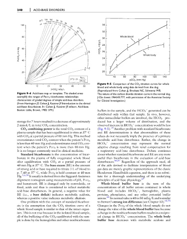
                                                                 Figure 9-5 Comparison of the CO 2 titration curves for whole
                        12      24      36      48     60        blood and whole body using data derived from the dog.
                                      –
                                  [HCO ] mEq/L                   (Reproduced from Cohen JJ, Brackett NC, Schwartz WB.
                                      3
            Figure 9-4 Acid-base map or template. The shaded areas  The nature of the carbon dioxide titration curve in the normal dog.
            exemplify the ranges of PaCO 2 -bicarbonate relationships  J Clin Invest 1964;43:777, with permission of the American Society
            characteristic of graded degrees of simple acid-base disorders.  for Clinical Investigation.)
            (From Harrington JT, Cohen JJ, Kassirer JP. Introduction to the clinical
            acid-base disturbances. In: Cohen JJ, Kassirer JP, editors. Acid-base.
            Boston: Little, Brown, 1982: 379.)                   buffers in the sample, and the HCO 3 generated can be

                                                                 distributed only within that sample. In vivo, however,

                                                                 other intracellular buffers are involved, the HCO 3 pro-
            storage for 7 hours resulted in a decrease of approximately  duced has a larger volume of distribution, and the
            2 mmol/L in total CO 2 concentration.                observed increase in HCO 3 concentration would be less

               CO 2 combining power is the total CO 2 content of a  (Fig. 9-5). 14  Another problem with standard bicarbonate
            plasma sample that has been equilibrated in vitro at 37 C  and BE determinations is that abnormalities of these

            with CO 2 at a partial pressure of 40 mm Hg. This method  values do not necessarily imply the presence of a primary
                                                                 metabolic acid-base disturbance. Rather, the change in
            overestimates total CO 2 content when the patient’s PCO 2
            is less than 40 mm Hg and underestimates total CO 2 con-  HCO 3    concentration may represent the normal
            tent when the patient’s PCO 2 is more than 40 mm Hg.  adaptive change resulting from renal compensation for
            It is no longer commonly used in clinical medicine.  a respiratory acid-base disturbance. Debate continues
               Standard bicarbonate is the concentration of bicar-  about whether standard bicarbonate and BE are any more
            bonate in the plasma of fully oxygenated whole blood  useful than bicarbonate in the evaluation of acid-base
            after equilibration with CO 2 at a partial pressure of  disturbances. 56,57  Regardless of the approach used, all

            40 mm Hg at 37 C. The base excess (BE) is the amount  of the aids devised to facilitate interpretation of blood
            of strong acid or base required to titrate 1 L of blood to  gas data are merely graphic representations of the classic
            p. 7.40 at 37 C, while PCO 2 is held constant at 40 mm  Henderson-Hasselbalch equation, and there is no substi-

            Hg. 5,6,59  It usually is derived from the Siggaard-Andersen  tute for a thorough understanding of the underlying
            alignment nomogram using measurements of pH, PCO 2 ,  principles of acid-base physiology. 67
            and hematocrit. BE is changed only by nonvolatile, or  Whole-blood buffer base is the sum of the
            fixed, acids and thus is considered to reflect metabolic  concentrations of all buffer anions contained in whole

            acid-base disturbances. In general, a negative value for  blood and includes HCO 3 , hemoglobin, plasma
            BE (i.e., a base deficit) indicates metabolic acidosis,  proteins, phosphates, and any other potential buffer
                                                                       60
            whereas a positive value indicates metabolic alkalosis.  anions. Itsnormalvalue is40to50 mEq/L andissimilar
               One problem with the concept of standard bicarbon-  to Stewart’s strong ion difference (see Chapter 13). 63,75
            ate is the assumption that the CO 2 titration curve of a  Changes in the PCO 2 of the whole blood sample do not
            whole blood sample is similar to that of the intact organ-  change the value of the whole blood buffer base because
            ism. This is not true because in the isolated blood sample,  achange in the nonbicarbonate buffers results in a recipro-

            all of the buffering of the CO 2 equilibrated with the sam-  cal change in HCO 3 concentration. The whole body
            ple is done by the hemoglobin and other nonbicarbonate  buffer base decreases with metabolic acidosis and

