Page 251 - Fluid, Electrolyte, and Acid-Base Disorders in Small Animal Practice
P. 251
242        ACID-BASE DISORDERS


            to establish a normal range because hyperventilation  they were the only samples not collected under free-
            related to fear or pain, increased muscular activity related  flowing conditions. Data for the normal dogs in this
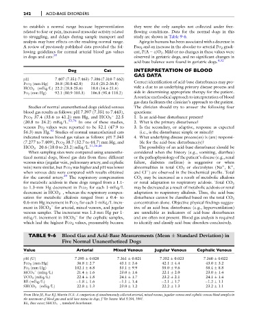
            to struggling, and delays during sample transport and  study are shown in Table 9-6.
            analysis may have effects on the resulting normal range.  Aging in humans has been associated with a decrease in
            A review of previously published data provided the fol-  Pao 2 and an increase in the alveolar-to-arterial PO 2 gradi-
            lowing guidelines for normal arterial blood gas values  ent, P(A   a)O 2 . Mild or no changes in these values were
            in dogs and cats: 25                                 observed in geriatric dogs, and no significant changes in
                                                                 acid-base balance were found in geriatric dogs. 4,32
                                 Dog               Cat           INTERPRETATION OF BLOOD
                                                                 GAS DATA
            pH             7.407 (7.351-7.463) 7.386 (7.310-7.462)
                                                                 Correct identification of acid-base disturbances may pro-
            PCO 2 (mm Hg)  36.8 (30.8-42.8)  31.0 (25.2-36.8)

            HCO 3 (mEq/L) 22.2 (18.8-25.6)  18.0 (14.4-21.6)     vide a clue to an underlying primary disease process and
            PO 2 (mm Hg)   92.1 (80.9-103.3)  106.8 (95.4-118.2)  aids in determining appropriate therapy for the patient.
                                                                 A routine methodical approach to interpretation of blood
                                                                 gas data facilitates the clinician’s approach to the patient.
               Studies of normal unanesthetized dogs yielded venous  The clinician should try to answer the following four
            blood gas results as follows: pH 7.397 (7.351 to 7.443),  questions:

            PCO 2 37.4 (33.6 to 41.2) mm Hg, and HCO 3  22.5     1. Is an acid-base disturbance present?
            (20.8 to 24.2) mEq/L. 52,76  In one of these studies,  2. What is the primary disturbance?
            venous PO 2 values were reported to be 52.1 (47.9 to  3. Is the secondary, or adaptive, response as expected
            56.3) mm Hg. 52  Studies of normal unanesthetized cats  (i.e., is the disturbance simple or mixed)?
            indicated venous blood gas values as follows: pH 7.343  4. What underlying disease process(es) is (are) responsi-
            (7.277 to 7.409), PCO 2 38.7 (32.7 to 44.7) mm Hg, and  ble for the acid-base disturbance(s)?
            HCO 3 20.6 (18.0 to 23.2) mEq/L. 11,26,46              The possibility of an acid-base disturbance should be

               When sampling sites were compared using unanesthe-  considered when the history (e.g., vomiting, diarrhea)
            tized normal dogs, blood gas data from three different  or the pathophysiology of the patient’s disease (e.g., renal
            venous sites (jugular vein, pulmonary artery, and cephalic  failure, diabetes mellitus) is suggestive or when
                                                                                                           þ
            vein) were similar, but PCO 2 was higher and pH was lower  abnormalities in total CO 2 or electrolytes (Na ,K ,
                                                                                                               þ
            when venous data were compared with results obtained  and Cl ) are observed in the biochemical profile. Total

            for the carotid artery. 29  The respiratory compensation  CO 2 may be increased as a result of metabolic alkalosis
            for metabolic acidosis in these dogs ranged from a 1.1-  or renal adaptation to respiratory acidosis. Total CO 2
            to 1.3-mm Hg decrement in PCO 2 for each 1-mEq/L     may be decreased as a result of metabolic acidosis or renal

            decrement in HCO 3 , whereas the respiratory compen-  adaptation to respiratory alkalosis. Thus, the acid-base
            sation for metabolic alkalosis ranged from a 0.4- to  disturbance cannot be classified based on the total CO 2
            0.6-mm Hg increment in PCO 2 for each 1-mEq/L incre-  concentration alone. Objective physical findings sugges-
            ment in HCO 3 for arterial, mixed venous, and jugular  tive of an acid-base disturbance (e.g., hyperventilation)

            venous samples. The increment was 1.3 mm Hg per 1-   are unreliable as indicators of acid-base disturbances

            mEq/L increment in HCO 3 for the cephalic samples,   and are often not present. Blood gas analysis is required
            which had the highest PCO 2 values, presumably because  to identify and classify acid-base disorders conclusively.

               TABLE 9-6      Blood Gas and Acid-Base Measurements (Mean   Standard Deviation) in
                              Five Normal Unanesthetized Dogs

            Value                    Arterial          Mixed Venous        Jugular Venous      Cephalic Venous
            pH (U)                   7.395   0.028       7.361   0.021       7.352   0.023        7.360   0.022
            PCO 2 (mm Hg)             36.8   2.7          43.1   3.6          42.1   4.4           43.0   3.2
            PO 2 (mm Hg)             102.1   6.8          53.1   9.9          55.0   9.6           58.4   8.8

            HCO 3 (mEq/L)             21.4   1.6          23.0   1.6          22.1   2.0           23.0   1.4
            TCO 2 (mEq/L)             22.4   1.8          24.1   1.7          23.2   2.1           24.1   1.4
            BE (mEq/L)                1.8   1.6            1.1   1.4           2.1   1.7            1.2   1.1

            SHCO 3 (mEq/L)            22.8   1.3          23.0   1.2          22.2   1.3           23.2   1.1
            From Ilkiw JE, Rose RJ, Martin ICA: A comparison of simultaneously collected arterial, mixed venous, jugular venous and cephalic venous blood samplesin
            the assessment of blood gas and acid base status in dogs, J Vet Intern Med 5:294, 1991.
            BE, Base excess; SHCO 3  , standard bicarbonate.

