
Page 57 - Fluid, Electrolyte, and Acid-Base Disorders in Small Animal Practice
P. 57
Disorders of Sodium and Water: Hypernatremia and Hyponatremia 47
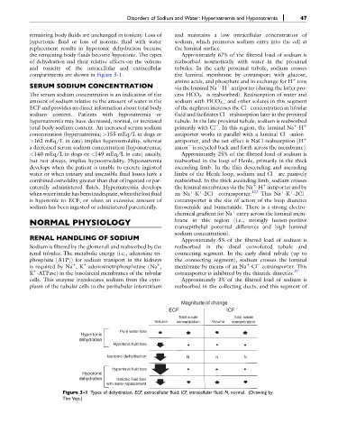
remaining body fluids are unchanged in tonicity. Loss of and maintains a low intracellular concentration of
hypertonic fluid or loss of isotonic fluid with water sodium, which promotes sodium entry into the cell at
replacement results in hypotonic dehydration because the luminal surface.
the remaining body fluids become hypotonic. The types Approximately 67% of the filtered load of sodium is
of dehydration and their relative effects on the volume reabsorbed isosmotically with water in the proximal
and tonicity of the intracellular and extracellular tubules. In the early proximal tubule, sodium crosses
compartments are shown in Figure 3-1. the luminal membrane by cotransport with glucose,
þ
amino acids, and phosphate and in exchange for H ions
SERUM SODIUM CONCENTRATION via the luminal Na -H antiporter (during the latter pro-
þ
þ
The serum sodium concentration is an indication of the cess HCO 3 is reabsorbed). Reabsorption of water and
amount of sodium relative to the amount of water in the sodium with HCO 3 and other solutes in this segment
ECF and provides no direct information about total body of the nephron increases the Cl concentration in tubular
sodium content. Patients with hyponatremia or fluid and facilitates Cl reabsorption later in the proximal
hypernatremia may have decreased, normal, or increased tubule. In the late proximal tubule, sodium is reabsorbed
total body sodium content. An increased serum sodium primarily with Cl . In this region, the luminal Na -H þ
þ
concentration (hypernatremia; >155 mEq/L in dogs or antiporter works in parallel with a luminal Cl -anion
>162 mEq/L in cats) implies hyperosmolality, whereas antiporter, and the net effect is NaCl reabsorption (H þ
a decreased serum sodium concentration (hyponatremia; anion is recycled back and forth across the membrane).
<140 mEq/L in dogs or <149 mEq/L in cats) usually, Approximately 25% of the filtered load of sodium is
but not always, implies hypoosmolality. Hyponatremia reabsorbed in the loop of Henle, primarily in the thick
develops when the patient is unable to excrete ingested ascending limb. In the thin descending and ascending
water or when urinary and insensible fluid losses have a limbs of the Henle loop, sodium and Cl are passively
combined osmolality greater than that of ingested or par- reabsorbed. In the thick ascending limb, sodium crosses
þ
enterally administered fluids. Hypernatremia develops the luminal membranes via the Na -H antiporter and by
þ
þ
þ
þ
whenwaterintakehasbeeninadequate, whenthe lost fluid an Na -K -2Cl cotransporter. 123 This Na -K -2Cl
þ
is hypotonic to ECF, or when an excessive amount of cotransporter is the site of action of the loop diuretics
sodium has been ingested or administered parenterally. furosemide and bumetanide. There is a strong electro-
chemical gradient for Na entry across the luminal mem-
þ
NORMAL PHYSIOLOGY brane in this region (i.e., strongly lumen-positive
transepithelial potential difference and high luminal
sodium concentration).
RENAL HANDLING OF SODIUM Approximately 5% of the filtered load of sodium is
Sodium is filtered by the glomeruli and reabsorbed by the reabsorbed in the distal convoluted tubule and
renal tubules. The metabolic energy (i.e., adenosine tri- connecting segment. In the early distal tubule (up to
phosphate [ATP]) for sodium transport in the kidneys the connecting segment), sodium crosses the luminal
þ
is required by Na ,K -adenosinetriphosphatase (Na , membrane by means of an Na -Cl cotransporter. This
þ
þ
þ
þ
K -ATPase) in the basolateral membranes of the tubular cotransporter is inhibited by the thiazide diuretics. 37
cells. This enzyme translocates sodium from the cyto- Approximately 3% of the filtered load of sodium is
plasm of the tubular cells to the peritubular interstitium reabsorbed in the collecting ducts, and this segment of
Magnitude of change
ECF ICF
Total solute Total solute
Volume concentration Volume concentration
Pure water loss
Hypertonic
dehydration
Hypotonic fluid loss
Isotonic dehydration N N N
Hypertonic fluid loss
Hypotonic
dehydration Isotonic fluid loss
with water replacement
Figure 3-1 Types of dehydration. ECF, extracellular fluid; ICF, intracellular fluid; N, normal. (Drawing by
Tim Vojt.)