
Page 62 - Fluid, Electrolyte, and Acid-Base Disorders in Small Animal Practice
P. 62
52 ELECTROLYTE DISORDERS
osmoreceptors and volume receptors. The volume of sodium and water from the proximal tubules mediated
receptors for the thirst mechanism are stimulated by by aquaporin 1 (AQP1) channels in the luminal and
angiotensin II and may be under control of the renin- basolateral membranes of these cells. In the presence of
angiotensin system. 108 volume depletion, RPF is usually decreased more than
The next most important stimulus for vasopressin the GFR, and enhanced proximal tubular reabsorption
release is volume depletion sensed by baroreceptors in of sodium and water may result from changes in
the left atrium, aortic sinus, and carotid sinuses. A postglomerular hemodynamics (see Fig. 3-3). These
decrease in blood volume of 5% to 10% lowers the thresh- factors may prevent adequate distal delivery of tubular
old for vasopressin release and increases the sensitivity fluid for dilution.
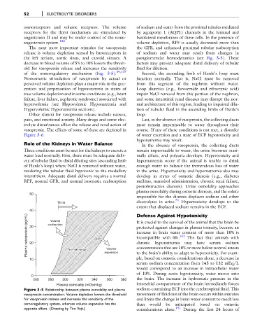
of the osmoregulatory mechanism (Fig. 3-5). 59,137 Second, the ascending limb of Henle’s loop must
Nonosmotic stimulation of vasopressin by actual or function normally. That is, NaCl must be removed
perceived volume depletion plays a major role in the gen- from this segment of the nephron without water.
eration and perpetuation of hyponatremia in states of Loop diuretics (e.g., furosemide and ethacrynic acid)
true volume depletion and in some conditions (e.g., heart impair NaCl removal from this portion of the nephron,
failure, liver failure, nephrotic syndrome) associated with and some interstitial renal diseases may disrupt the nor-
hypervolemia (see Hypovolemic Hyponatremia and mal architecture of this region, leading to impaired dilu-
Hypervolemic Hyponatremia sections). tion of tubular fluid in the ascending limbs of Henle’s
Other stimuli for vasopressin release include nausea, loop.
pain, and emotional anxiety. Many drugs and some elec- Last, in the absence of vasopressin, the collecting ducts
trolyte disturbances affect the release and renal action of must remain impermeable to water throughout their
vasopressin. The effects of some of these are depicted in course. If any of these conditions is not met, a disorder
Figure 3-6. of water excretion and a state of ECF hypotonicity and
hyponatremia may result.
Role of the Kidneys in Water Balance In the absence of vasopressin, the collecting ducts
Three conditions must be met for the kidneys to excrete a remain impermeable to water, the urine becomes maxi-
water load normally. First, there must be adequate deliv- mally dilute, and polyuria develops. Hypertonicity and
ery of tubular fluid to distal diluting sites (ascending limb hypernatremia occur if the animal is unable to drink
of Henle’s loop) where NaCl is removed without water, enough water to balance the tremendous loss of water
rendering the tubular fluid hypotonic to the medullary in the urine. Hypertonicity and hypernatremia also may
interstitium. Adequate distal delivery requires a normal develop in states of osmotic diuresis (e.g., diabetes
RPF, normal GFR, and normal isosmotic reabsorption mellitus, mannitol administration, chronic renal failure,
postobstructive diuresis). Urine osmolality approaches
plasma osmolality during osmotic diuresis, and the solute
50
responsible for the diuresis displaces sodium and other
Thirst electrolytes in urine. 51 Hypertonicity develops to the
extent that displaced sodium remains in the ECF.
40 Volume Defense Against Hypotonicity
Plasma vasopressin (pg/mL) 30 contraction protected against changes in plasma tonicity, because an
It is crucial to the survival of the animal that the brain be
increase in brain water content of more than 10% is
151
The fact that animals with
incompatible with life.
have
may
chronic
hyponatremia
sodium
serum
20
Volume
to the brain’s ability to adapt to hypotonicity. For exam-
ple, based on osmotic considerations alone, a decrease in
10 Normal range expansion concentrations that are 10% or more below normal attests
serum sodium concentration from 145 to 132 mEq/L
would correspond to an increase in intracellular water
of 10%. During acute hypotonicity, water moves into
0
260 280 300 320 340 360 380 the brain. The increase in hydrostatic pressure in the
Plasma osmolality (mOsm/kg) interstitial compartment of the brain immediately forces
Figure 3-5 Relationship between plasma osmolality and plasma sodium-containing ECF into the cerebrospinal fluid. This
vasopressin concentration. Volume depletion lowers the threshold movement of fluid out of the brain occurs within minutes
for vasopressin release and increases the sensitivity of the and limits the change in brain water content to much less
osmoregulatory system, whereas volume expansion has the than would be anticipated based on osmotic
opposite effect. (Drawing by Tim Vojt.) considerations alone. 151 During the first 24 hours of