
Page 71 - Fluid, Electrolyte, and Acid-Base Disorders in Small Animal Practice
P. 71
Disorders of Sodium and Water: Hypernatremia and Hyponatremia 61
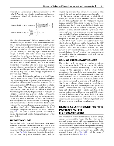
presentation, and its serum sodium concentration is 170 original replacement fluid should be isotonic so that
mEq/L.Assuminga previously normal serumsodium con- extracellular volume repletion can proceed rapidly.
centration of 145 mEq/L, the dog’s water deficit can be In the presence of hemorrhagic shock, whole blood,
calculated: plasma, or a colloid solution is the ideal fluid to adminis-
ter. The hemoglobin in whole blood improves oxygen-
carrying capacity. The plasma proteins in whole blood
0 1
P Na ðpresentÞ
Water deficit ¼ WtðpresentÞ @ 1 A and plasma or the dextrans in a colloid solution increase
P Na ðpreviousÞ
and maintain intravascular volume by increasing oncotic
pressure. In many animals that have experienced severe
0 1
170 hypotonic losses over an extended time period, replace-
Water deficit ¼ 10 @ 1 A ¼ 1:72 L
145 ment of the ECF volume with an isotonic crystalloid solu-
tion (e.g., 0.9% NaCl and lactated Ringer’s solution) is
adequate. A volume up to four times the suspected intra-
The original estimates of TBW and serum sodium con- vascular deficit may be required because the isotonic crys-
centration may be modified based on information avail- talloid solution distributes rapidly throughout the ECF
able to the clinician at presentation. For example, if the compartment (ECF volume is four times intravascular
dog’s normal serum sodium concentration is known from volume). After the extracellular volume has been
a previous admission, this value can be substituted in expanded, hypotonic fluids (e.g., 0.45% NaCl and half-
place of 145 mEq/L. If the dog’s previous normal body strength lactated Ringer’s solution) can be administered
weight is known, the water deficit may simply be to provide fluids for maintenance needs and ongoing
estimated as the difference between the previous and losses (see Chapter 14).
present body weights. The assumption inherent in the lat-
ter calculation is that the patient has not gained or lost tis- GAIN OF IMPERMEANT SOLUTE
sue mass. For a short period, this is a reasonable The patient with an excess of sodium-containing
assumption because loss of 1 kg of tissue mass requires impermeant solute in the ECF can be treated by admin-
an expenditure of approximately 1600 kcal. This caloric istration of 5% dextrose intravenously. The main disad-
expenditure would require fasting for 2 to 3 days in a nor- vantage of this approach is that it causes further
mal 10-kg dog with a basal energy requirement of expansion of the extracellular compartment in a patient
approximately 700 kcal. already suffering from ECF volume expansion. In an ani-
A pure water deficit can be replaced by giving 5% dex- mal with normal cardiac and renal function, this volume
trose in water intravenously. This solution technically is expansion leads to diuresis and natriuresis, and ECF vol-
only slightly hypotonic to plasma (278 mOsm/kg), but ume returns to normal. In an animal with underlying car-
the glucose ultimately enters cells and is metabolized so diac disease or oliguria related to primary renal disease,
that administration of 5% dextrose is equivalent to admin- this approach may lead to development of pulmonary
istration of water. The water deficit must be replaced and edema. Administration of a loop diuretic (e.g., furose-
hypernatremia corrected slowly over 48 hours. The brain mide and ethacrynic acid) promotes excretion of the
adapts to hypertonicity by the production of osmolytes or existing sodium load and hastens return of ECF volume
idiogenic osmoles that prevent cellular dehydration. to normal. As in the case of pure water deficit, it is essen-
Excessively rapid lowering of the serum sodium concen- tial that fluid administration proceeds slowly and that
tration may result in movement of water into brain cells serum sodium concentration be lowered gradually over
and development of cerebral edema. In human patients 48 hours to avoid neurologic complications.
with hypernatremia of chronic or unknown duration, cor-
rection of the serum sodium concentration at a rate of less
than 10 to 12 mEq/L per 24 hours minimizes the risk of CLINICAL APPROACH TO THE
neurologic complications related to water intoxica- PATIENT WITH
tion. 6,152 The animal’s serum sodium concentration HYPONATREMIA
should be monitored serially during replacement of the
water deficit. The presence of hyponatremia usually, but not always,
implies hypoosmolality. Thus, the first step in the
HYPOTONIC LOSS approach to the patient with hyponatremia is to deter-
As described earlier, hypotonic losses cause more severe mine whether hypoosmolality of the ECF is present. This
extracellular volume contraction than do losses of pure can be determined by measurement of plasma osmolality.
water. As the tonicity of the fluid lost approaches the The evaluation of hyponatremia then may be approached
tonicity of ECF, the extracellular volume deficit becomes using the patient’s plasma osmolality as a guide. This
greater (see Fig. 3-9). As a result, signs of volume deple- approach is outlined in Fig. 3-12, and the causes of
tion are more likely with hypotonic losses, and the hyponatremia are listed in Box 3-3.