
Page 68 - Fluid, Electrolyte, and Acid-Base Disorders in Small Animal Practice
P. 68
58 ELECTROLYTE DISORDERS
weak pulses, and delayed capillary refill time). As the tonicity, the greater the volume loss from the ECF
tonicity of the fluid lost increases toward the normal compartment.
tonicity of ECF, the volume deficit of the extracellular For simplicity, these examples are based on many
compartment becomes progressively more severe assumptions that in reality may not be true. For example,
(Fig. 3-9). In the case of isotonic losses, no osmotic stim- TBW is not 60% of body weight in all individuals, the
ulus for water movement is present. The entire loss is number of osmoles in the ECF may have been altered
borne by the extracellular compartment, and by electrolyte losses not detected clinically, the effects
hypovolemic shock may occur if the loss has been of suf- of hydrostatic forces resulting from extracellular volume
ficient magnitude (e.g., severe hemorrhage). depletion have not been considered, some solutes may
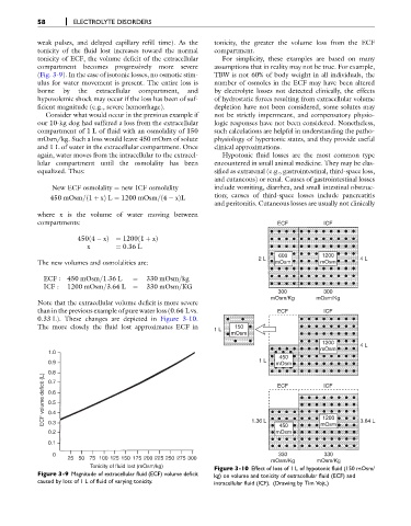
Consider what would occur in the previous example if not be strictly impermeant, and compensatory physio-
our 10-kg dog had suffered a loss from the extracellular logic responses have not been considered. Nonetheless,
compartment of 1 L of fluid with an osmolality of 150 such calculations are helpful in understanding the patho-
mOsm/kg. Such a loss would leave 450 mOsm of solute physiology of hypertonic states, and they provide useful
and 1 L of water in the extracellular compartment. Once clinical approximations.
again, water moves from the intracellular to the extracel- Hypotonic fluid losses are the most common type
lular compartment until the osmolality has been encountered in small animal medicine. They may be clas-
equalized. Thus: sified as extrarenal (e.g., gastrointestinal, third-space loss,
and cutaneous) or renal. Causes of gastrointestinal losses
New ECF osmolality ¼ new ICF osmolality include vomiting, diarrhea, and small intestinal obstruc-
450 mOsm= 1 þ xÞ L ¼ 1200 mOsm=ð4 xÞL tion; causes of third-space losses include pancreatitis
ð
and peritonitis. Cutaneous losses are usually not clinically
where x is the volume of water moving between
compartments: ECF ICF
450ð4 xÞ¼ 1200ð1 þ xÞ
x ¼ 0:36 L
600 1200
2 L 4 L
The new volumes and osmolalities are: mOsm mOsm
ECF : 450 mOsm=1:36 L ¼ 330 mOsm=kg
ICF : 1200 mOsm=3:64 L ¼ 330 mOsm=KG
300 300
mOsm/Kg mOsm/Kg
Note that the extracellular volume deficit is more severe
than in the previous example of pure water loss (0.64 L vs. ECF ICF
0.33 L). These changes are depicted in Figure 3-10.
The more closely the fluid lost approximates ECF in 1 L 150
mOsm
1200
mOsm 4 L
1.0
450
0.9 1 L mOsm
0.8 ECF ICF
ECF volume deficit (L) 0.6
0.7
0.5
0.4
1200
0.3
450
0.2 1.36 L mOsm mOsm 3.64 L
0.1
0 330 330
25 50 75 100 125 150 175 200 225 250 275 300
mOsm/Kg mOsm/Kg
Tonicity of fluid lost (mOsm/kg) Figure 3-10 Effect of loss of 1 L of hypotonic fluid (150 mOsm/
Figure 3-9 Magnitude of extracellular fluid (ECF) volume deficit kg) on volume and tonicity of extracellular fluid (ECF) and
caused by loss of 1 L of fluid of varying tonicity. intracellular fluid (ICF). (Drawing by Tim Vojt.)