
Page 129 - The Toxicology of Fishes
P. 129
Toxicokinetics in Fishes 109
VASCULAR
Q i Q i
SPACE
C art C vi
k i
EXTRAVASCULAR
SPACE
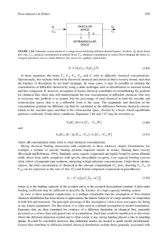
FIGURE 3.24 Schematic representation of a compartment exhibiting diffusion-limited kinetics. Symbols: Q i , tissue blood
flow rate; C art , chemical concentration in arterial blood; C vi , chemical concentration in venous blood draining the tissue; k i ,
transport parameter used to model diffusive flux across the capillary endothelium.
C i = ( V C bi + V C ei) V i (3.89)
ei
bi
In these equations, the terms C , C , C , C , and C refer to diffusible chemical concentrations.
i
bi
ei
a
vi
Operationally, this includes both freely dissolved chemical and chemical that is loosely bound, such that
the kinetics of desorption do not limit exchange. In some cases, it may be possible to estimate the
concentration of diffusible chemical by using a static technique such as ultrafiltration to measure bound
and free compound. If, however, desorption of bound chemical contributes to reestablishing the gradient
for chemical flux, these data will underestimate the true concentration of diffusible chemical. One way
to overcome this problem is to assume that the percentage of total chemical in both the vascular and
extravascular spaces that is in a diffusible form is the same. The magnitude and direction of the
concentration gradient for diffusion can then be calculated as the difference between chemical concen-
tration in the vascular space and that in the extravascular space, divided by a tissue–blood equilibrium
partition coefficient. Under these conditions, Equations 3.86 and 3.87 may be rewritten as:
VdC dt = ( C P i) (3.90)
k C vi −
ei
ei
i
i
and
dt = ( C vi) − ( C P i)
k C vi −
Q C art −
bi
VdC bi i i ei (3.91)
where all concentration terms refer to total chemical concentrations.
Strong chemical binding interactions add complexity to these relatively simple formulations; for
example, a number of specific binding proteins sequester metals in tissues, limiting their toxicity
(Roesijadi and Robinson, 1994). Similarly, some organic compounds are highly bound by serum albumin
while others form stable complexes with specific intracellular receptors. Low-capacity binding systems
often follow a Langmuir-type isotherm, saturating at high substrate concentrations. Under these circum-
stances, the total concentration of chemical in the relevant compartment (C ; here, denoting C , C , or
ei
i
j
*
C ) can be expressed as the sum of free (C ) and bound compound (expression in parentheses):
bi
j
C j = C j +( α i C j ( ε j + C j)) (3.92)
*
*
*
where α is the binding capacity of the receptor and ε is the receptor dissociation constant. A first-order
j
j
binding coefficient may be sufficient to describe the kinetics of a high-capacity binding system.
In view of these potential complexities, it is perhaps remarkable that the assumption of flow-limited
chemical distribution has been shown to approximate the kinetic behavior of a large number of compounds
in both fish and mammals. The principal advantage of this assumption is that it does not require the fitting
of any kinetic parameters. For this reason, it is often used as a default assumption in model formulation.
Exposure data are then examined for evidence of a diffusion limitation on chemical flux, generally
presented as a slower than anticipated rate of accumulation. Such data would be insufficient to determine
where the diffusion limitation existed and to what extent, if any, strong binding played a role in retarding
uptake. It could be concluded, however, that additional studies are needed to guide model development.
Factors that contribute to diffusion-limited chemical distribution include those generally associated with