
Page 424 - The Toxicology of Fishes
P. 424
404 The Toxicology of Fishes
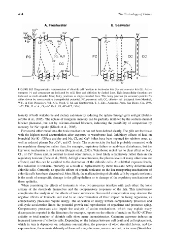
A. Freshwater B. Seawater
Ca 2+
Na + CI – + CI – Na + +
Na Na
H + Ca 2+ HCO 3 – NH 4 + Ca 2+
H +
–
PC CO 2 H 2 O PC CI K + CC CC
CI – Na +
CC
K +
Ca 2+ Ca 2+ 2K + 3Na + Ca 2+ Ca 2+ 2K + 3Na +
~ ~ ~ ~
–
3Na + 2H + 3Na + 2H + 2CI Na +
K +
FIGURE 8.2 Diagrammatic representation of chloride cell function in freshwater fish (A) and seawater fish (B). Active
transport (~) and cotransport are indicated by solid lines and diffusion by dashed lines. Tight intercellular functions are
indicated as multi-stranded lines; leaky junctions as single-stranded lines. This leaky junction (in seawater) permits Na
efflux driven by serosa-positive transepithelial potential. PC, pavement cell; CC, chloride cell. (Adapted from Marshall,
W.S., in Fish Physiology, Vol. XIV, Wood, C. M. and Shuttleworth, T. J., Eds., Academic Press, San Diego, CA, 1995,
1–23; Flik, G. et al., Physiol. Zool., 69, 403–417, 1996.)
toxicity of both waterborne and dietary cadmium by reducing the uptake through gills and gut (Baldis-
serotto et al., 2005). The uptake of inorganic mercury can be partially inhibited by the sodium-channel
blocker phenamel, but not by calcium-channel blockers, indicating the possibility of competition by
+
mercury for Na uptake (Klinck et al., 2005).
For several other metal ions, the toxic mechanism has not been defined clearly. The gills are the tissue
with the highest metal accumulation after exposure to waterborne lead. Inhibitory effects of lead on
branchial Na /K -ATPase activity and Na, Cl, and Ca influx have been reported for rainbow trout, as
+
2+
+
2+
+
–
well as reduced plasma Na , Ca , and Cl levels. The acute toxicity for lead is probably connected with
ion regulatory disruption rather than, for example, respiratory failure or acid–base disturbance, but the
+
key toxic mechanism is still unclear (Rogers et al., 2003). Waterborne nickel has no clear effect on Na ,
Cl , or Ca fluxes and, in contrast to most other metals, is most likely a respiratory rather than an ion
–
2+
regulatory toxicant (Pane et al., 2003). At high concentrations, the plasma levels of many other ions are
affected, and this can be ascribed to the destruction of the chloride cells. At sublethal exposure levels,
this reduction is transient, probably as a result of replacement by more resistant newly differentiated
chloride cells. Currently, no specific effects of organic toxicants on the ion-transporting mechanisms of
chloride cells have been determined. Most likely, the malfunctioning of chloride cells by organic toxicants
is the result of nonspecific damage to the gill epithelium or to damage of the regulatory mechanisms of
these epithelia.
When examining the effects of toxicants in vivo, two processes interfere with each other: the toxic
actions of the chemicals themselves and the compensatory responses of the fish. This interference
complicates the analysis of the effects of toxic substances. Successful compensation may obscure the
negative effects of toxicants and lead to an underestimation of their impact on living organisms, as
compensatory processes require energy. The allocation of energy toward compensatory processes and
cell-cycle acceleration limits the potential growth and reproduction of organisms and promotes aging.
Compensatory processes also impair the analysis of action mechanisms, which may explain many
+
+
discrepancies reported in the literature; for example, reports on the effects of metals on Na /K -ATPase
activity or total number of chloride cells show many inconsistencies. Cadmium exposure induces an
increased turnover of chloride cells. Depending on the balance between cell death and cell replacement,
which in turn is dependent on cadmium concentration, the presence of other stressful factors, and the
exposure time, the numerical density of these cells may decrease, remain constant, or increase (Wendelaar