
Page 484 - The Toxicology of Fishes
P. 484
464 The Toxicology of Fishes
1 2
STEROID Membrane STEROID
Receptor
Cross-talk?
STEROID
3 fast
Nuclear
activates 2nd messengers
Receptor (often nongenomic)
4
DNA slow
Nucleus activates genes
(genomic)
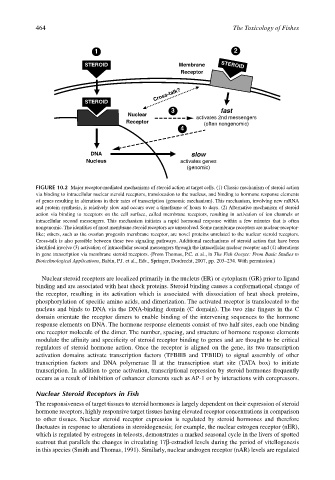
FIGURE 10.2 Major receptor-mediated mechanisms of steroid action at target cells. (1) Classic mechanism of steroid action
via binding to intracellular nuclear steroid receptors, translocation to the nucleus, and binding to hormone response elements
of genes resulting in alterations in their rates of transcription (genomic mechanism). This mechanism, involving new mRNA
and protein synthesis, is relatively slow and occurs over a timeframe of hours to days. (2) Alternative mechanism of steroid
action via binding to receptors on the cell surface, called membrane receptors, resulting in activation of ion channels or
intracellular second messengers. This mechanism initiates a rapid hormonal response within a few minutes that is often
nongenomic. The identities of most membrane steroid receptors are unresolved. Some membrane receptors are nuclear-receptor-
like; others, such as the ovarian progestin membrane receptor, are novel proteins unrelated to the nuclear steroid receptors.
Cross-talk is also possible between these two signaling pathways. Additional mechanisms of steroid action that have been
identified involve (3) activation of intracellular second messengers through the intracellular nuclear receptor and (4) alterations
in gene transcription via membrane steroid receptors. (From Thomas, P.C. et al., in The Fish Oocyte: From Basic Studies to
Biotechnological Applications, Babin, P.J. et al., Eds., Springer, Dordrecht, 2007, pp. 203–234. With permission.)
Nuclear steroid receptors are localized primarily in the nucleus (ER) or cytoplasm (GR) prior to ligand
binding and are associated with heat shock proteins. Steroid binding causes a conformational change of
the receptor, resulting in its activation which is associated with dissociation of heat shock proteins,
phosphorylation of specific amino acids, and dimerization. The activated receptor is translocated to the
nucleus and binds to DNA via the DNA-binding domain (C domain). The two zinc fingers in the C
domain orientate the receptor dimers to enable binding of the intervening sequences to the hormone
response elements on DNA. The hormone response elements consist of two half sites, each one binding
one receptor molecule of the dimer. The number, spacing, and structure of hormone response elements
modulate the affinity and specificity of steroid receptor binding to genes and are thought to be critical
regulators of steroid hormone action. Once the receptor is aligned on the gene, its two transcription
activation domains activate transcription factors (TFBIIB and TFBIID) to signal assembly of other
transcription factors and DNA polymerase II at the transcription start site (TATA box) to initiate
transcription. In addition to gene activation, transcriptional repression by steroid hormones frequently
occurs as a result of inhibition of enhancer elements such as AP-1 or by interactions with corepressors.
Nuclear Steroid Receptors in Fish
The responsiveness of target tissues to steroid hormones is largely dependent on their expression of steroid
hormone receptors, highly responsive target tissues having elevated receptor concentrations in comparison
to other tissues. Nuclear steroid receptor expression is regulated by steroid hormones and therefore
fluctuates in response to alterations in steroidogenesis; for example, the nuclear estrogen receptor (nER),
which is regulated by estrogens in teleosts, demonstrates a marked seasonal cycle in the livers of spotted
seatrout that parallels the changes in circulating 17β-estradiol levels during the period of vitellogenesis
in this species (Smith and Thomas, 1991). Similarly, nuclear androgen receptor (nAR) levels are regulated