
Page 511 - The Toxicology of Fishes
P. 511
The Immune System of Fish: A Target Organ of Toxicity 491
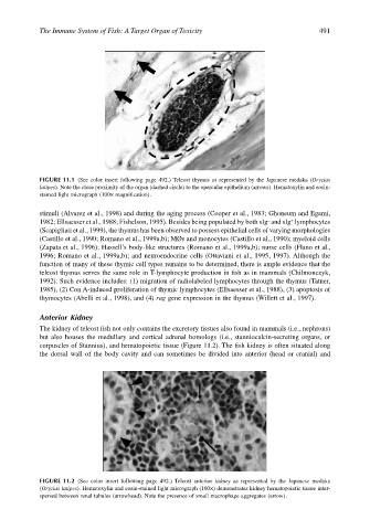
FIGURE 11.1 (See color insert following page 492.) Teleost thymus as represented by the Japanese medaka (Oryzias
latipes). Note the close proximity of the organ (dashed circle) to the opercular epithelium (arrows). Hematoxylin and eosin-
stained light micrograph (100× magnification).
stimuli (Alvarez et al., 1998) and during the aging process (Cooper et al., 1983; Ghoneum and Egami,
–
+
1982; Ellsaesser et al., 1988; Fishelson, 1995). Besides being populated by both sIg and sIg lymphocytes
(Scapigliati et al., 1999), the thymus has been observed to possess epithelial cells of varying morphologies
(Castillo et al., 1990; Romano et al., 1999a,b); MØs and monocytes (Castillo et al., 1990); myeloid cells
(Zapata et al., 1996); Hassell’s body-like structures (Romano et al., 1999a,b); nurse cells (Flano et al.,
1996; Romano et al., 1999a,b); and neuroendocrine cells (Ottaviani et al., 1995, 1997). Although the
function of many of these thymic cell types remains to be determined, there is ample evidence that the
teleost thymus serves the same role in T-lymphocyte production in fish as in mammals (Chilmonczyk,
1992). Such evidence includes: (1) migration of radiolabeled lymphocytes through the thymus (Tatner,
1985), (2) Con A-induced proliferation of thymic lymphocytes (Ellsaesser et al., 1988), (3) apoptosis of
thymocytes (Abelli et al., 1998), and (4) rag gene expression in the thymus (Willett et al., 1997).
Anterior Kidney
The kidney of teleost fish not only contains the excretory tissues also found in mammals (i.e., nephrons)
but also houses the medullary and cortical adrenal homologs (i.e., stanniocalcin-secreting organs, or
corpuscles of Stannius), and hematopoietic tissue (Figure 11.2). The fish kidney is often situated along
the dorsal wall of the body cavity and can sometimes be divided into anterior (head or cranial) and
FIGURE 11.2 (See color insert following page 492.) Teleost anterior kidney as represented by the Japanese medaka
(Oryzias latipes). Hematoxylin and eosin-stained light micrograph (100×) demonstrates kidney hematopoietic tissue inter-
spersed between renal tubules (arrowhead). Note the presence of small macrophage aggregates (arrow).