
Page 512 - The Toxicology of Fishes
P. 512
492 The Toxicology of Fishes
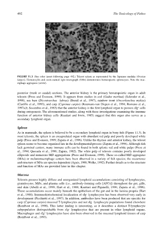
FIGURE 11.3 (See color insert following page 492.) Teleost spleen as represented by the Japanese medaka (Oryzias
latipes). Hematoxylin and eosin-stained light micrograph (100×) demonstrates hematopoetic splenocytes. Note the mac-
rophage aggregates (arrow).
posterior (trunk or caudal) sections. The anterior kidney is the primary hematopoietic organ in adult
teleosts (Press and Evensen, 1999). It appears from studies in cod (Gadus morhua) (Schroder et al.,
1998), sea bass (Dicentrarchus labrax) (Breuil et al., 1997), rainbow trout (Oncorhynchus mykiss)
(Castillo et al., 1993), and carp (Cyprinus carpio) (Koumans-van Diepen et al., 1994; Romano et al.,
+
1997a,b; Secombes et al., 1983) that the anterior kidney is the first lymphoid organ to possess sIg cells
during ontogenesis. The aforementioned studies, along with those investigations examining the immune
function of anterior kidney cells (Kaattari and Irwin, 1985) suggest that this organ also serves as a
secondary lymphoid organ.
Spleen
As in mammals, the spleen is believed to be a secondary lymphoid organ in bony fish (Figure 11.3). In
most teleosts, the spleen is an encapsulated organ with abundant red pulp and poorly developed white
pulp (Press and Evensen, 1999; Zapata et al., 1996). Unlike the thymus and anterior kidney, the teleost
spleen seems to become organized late in the developmental process (Zapata et al., 1996). Although fish
lack germinal centers, many immune cells can be found in both splenic red and white pulps (Press et
al., 1994; Quesada et al., 1990; Zapata, 1982). The white pulp of teleosts contains poorly developed
ellipsoids and numerous MØ aggregations (Press and Evensen, 1999). These so-called MØ aggregates
(MAs) or melanomacrophage centers have been observed in a variety of fish species; the occurrence
and structure of MAs are species dependent (Aguis, 1980; Wolke, 1992). Further details as to the structure
and function of MAs are provided later in this chapter.
Mucosa
Teleosts possess highly diffuse and unorganized lymphoid accumulations consisting of lymphocytes,
granulocytes, MØs, and plasma cells (i.e., antibody-forming cells [AFCs]) throughout the gut, gills,
and skin (Abelli et al., 1999; Hart et al., 1988; Kaattari and Piganelli, 1996; Zapata et al., 1996).
These accumulations occur mainly beneath the epithelium of the gut and in the lamina propria (Hart
–
et al., 1988). Immunohistochemical localization of sIg lymphocytes has been observed very early in
development (Picchietti et al., 1997). In addition, antibodies have been produced that are specific for
carp (Cyprinus carpio) mucosal T-lymphocytes and not sIg lymphocyte populations found elsewhere
–
(Rombout et al., 1998). This latter finding is interesting, as it describes a distinct T-lymphocyte
subpopulation distinguishable from sIg lymphocytes that are present in other lymphoid organs.
–
+
Macrophages and sIg lymphocytes have also been observed in the mucosal lymphoid tissues of carp
(Rombout et al., 1993).