Page 148 - Feline Cardiology
P. 148
Chapter 11: Hypertrophic Cardiomyopathy  147


                   5                                                 30
                                                                           Volume Derivative
                                                                           End Systole
                  4.5                                                20    MR EFR
                                                                           MR AFR
                   4                                                 10    End Diastole
                 Volume [cm 3 ]  3.5                Volume          dV/dt [cm 3 /s]  0


                   3                                End Systole     –10
                                                    End Diastole
                                                    MidPoint
                  2.5                                               –20


                   2                                                –30                                                 Cardiomyopathies
                    0    0.1   0.2   0.3  0.4   0.5   0.6   0.7       0     0.1  0.2   0.3   0.4  0.5   0.6   0.7
              A                      Time [sec]                  C                      Time [sec]

                  30
                                                Volume Derivative
                                                End Systole
                  20                            MR EFR
                                                MR AFR
                                                End Diastole
                  10
                 dV/dt [cm 3 /s]  0


                  –10


                  –20

                  –30
                    0    0.1   0.2   0.3  0.4   0.5   0.6  0.7
              B                      Time [sec]
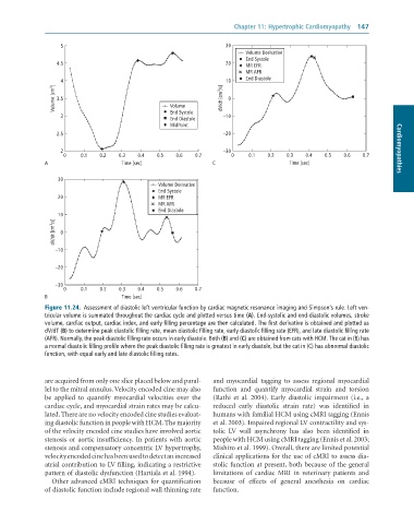
              Figure 11.24.  Assessment	of	diastolic	left	ventricular	function	by	cardiac	magnetic	resonance	imaging	and	Simpson’s	rule.	Left	ven-
              tricular	volume	is	summated	throughout	the	cardiac	cycle	and	plotted	versus	time	(A).	End-systolic	and	end-diastolic	volumes,	stroke
              volume,	cardiac	output,	cardiac	index,	and	early	filling	percentage	are	then	calculated.	The	first	derivative	is	obtained	and	plotted	as
              dV/dT	(B)	to	determine	peak	diastolic	filling	rate,	mean	diastolic	filling	rate,	early	diastolic	filling	rate	(EFR),	and	late	diastolic	filling	rate
              (AFR).	Normally,	the	peak	diastolic	filling	rate	occurs	in	early	diastole.	Both	(B)	and	(C)	are	obtained	from	cats	with	HCM.	The	cat	in	(B)	has
              a	normal	diastolic	filling	profile	where	the	peak	diastolic	filling	rate	is	greatest	in	early	diastole,	but	the	cat	in	(C)	has	abnormal	diastolic
              function,	with	equal	early	and	late	diastolic	filling	rates.


              are acquired from only one slice placed below and paral-  and  myocardial  tagging  to  assess  regional  myocardial
              lel to the mitral annulus. Velocity encoded cine may also   function  and  quantify  myocardial  strain  and  torsion
              be  applied  to  quantify  myocardial  velocities  over  the   (Rathi  et  al.  2004).  Early  diastolic  impairment  (i.e.,  a
              cardiac cycle, and myocardial strain rates may be calcu-  reduced  early  diastolic  strain  rate)  was  identified  in
              lated. There are no velocity encoded cine studies evaluat-  humans with familial HCM using cMRI tagging (Ennis
              ing diastolic function in people with HCM. The majority   et al. 2003). Impaired regional LV contractility and sys-
              of the velocity encoded cine studies have involved aortic   tolic  LV  wall  asynchrony  has  also  been  identified  in
              stenosis or aortic insufficiency. In patients with aortic   people with HCM using cMRI tagging (Ennis et al. 2003;
              stenosis and compensatory concentric LV hypertrophy,   Mishiro et al. 1999). Overall, there are limited potential
              velocity encoded cine has been used to detect an increased   clinical applications for the use of cMRI to assess dia-
              atrial contribution to LV filling, indicating a restrictive   stolic function at present, both because of the general
              pattern of diastolic dysfunction (Hartiala et al. 1994).  limitations  of  cardiac  MRI  in  veterinary  patients  and
                 Other advanced cMRI techniques for quantification   because  of  effects  of  general  anesthesia  on  cardiac
              of diastolic function include regional wall thinning rate   function.

