Page 188 - Feline Cardiology
P. 188
190  Section D: Cardiomyopathies


              et  al.  2000).  No  breed  predisposition  has  been  noted   first-degree AV block, and third-degree AV block may
              (Fox et al. 2000; Harvey et al. 2005).             be  observed  alone  or  in  combination  (see  Chapter
                                                                 18).
              HISTORY AND CHIEF COMPLAINT
                                                                 Radiography
              The majority of affected cats have presented for vague   Right  atrial  and  ventricular  enlargements  are  almost
              clinical signs, including lethargy and anorexia; addition-  always observed, and findings consistent with right heart
              ally, signs of right heart failure including dyspnea and   failure (pleural effusion, caudal caval distention) are fre-
              tachypnea may be observed. Syncope may be observed   quently present.
              but appears to be uncommon (Fox et al. 2000; Harvey   Echocardiography
              et  al.  2005).  Sudden  death  is  possible.  ARVC  may  be
      Cardiomyopathies  detected incidentally in cats not showing overt clinical   Echocardiography is needed to confirm the diagnosis.
              signs,  but  the  majority  of  cases  are  identified  due  to
                                                                 The significant right-sided lesions consist of right atrial
              presentation  related  to  overt  clinical  signs  (Fox  1999;
                                                                 and right ventricular enlargement and decreased right
              Paige et al. 2009).
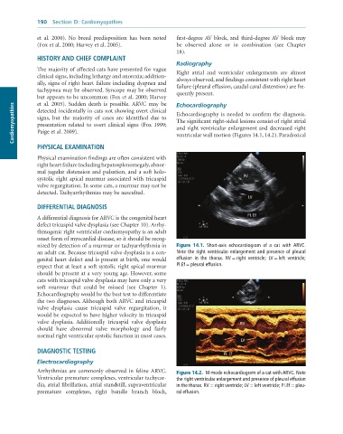
                                                                 ventricular wall motion (Figures 14.1, 14.2). Paradoxical
              PHYSICAL EXAMINATION

              Physical examination findings are often consistent with
              right heart failure including hepatosplenomegaly, abnor-
              mal jugular distension and pulsation, and a soft holo-
              systolic right apical murmur associated with tricuspid                     RV
              valve regurgitation. In some cats, a murmur may not be
              detected. Tachyarrhythmias may be ausculted.
                                                                                           LV
              DIFFERENTIAL DIAGNOSIS
              A differential diagnosis for ARVC is the congenital heart                    PI. Ef
              defect tricuspid valve dysplasia (see Chapter 10). Arrhy-
              thmogenic right ventricular cardiomyopathy is an adult
              onset form of myocardial disease, so it should be recog-
              nized by detection of a murmur or tachyarrhythmia in   Figure  14.1.  Short-axis	 echocardiogram	 of	 a	 cat	 with	 ARVC.
              an adult cat. Because tricuspid valve dysplasia is a con-  Note	the	right	ventricular	enlargement	and	presence	of	pleural
              genital heart defect and is present at birth, one would   effusion	 in	 the	 thorax.	 RV	=	right	 ventricle;	 LV	=	left	 ventricle;
              expect that at least a soft systolic right apical murmur   Pl.Ef	=	pleural	effusion.
              should be present at a very young age. However, some
              cats with tricuspid valve dysplasia may have only a very
              soft  murmur  that  could  be  missed  (see  Chapter  1).
              Echocardiography would be the best test to differentiate
              the two diagnoses. Although both ARVC and tricuspid
              valve  dysplasia  cause  tricuspid  valve  regurgitation,  it
              would be expected to have higher velocity in tricuspid
              valve  dysplasia.  Additionally  tricuspid  valve  dysplasia              RV
              should  have  abnormal  valve  morphology  and  fairly
              normal right ventricular systolic function in most cases.
                                                                                        LV
              DIAGNOSTIC TESTING
                                                                                  PI. Ef
              Electrocardiography
              Arrhythmias  are  commonly  observed  in  feline ARVC.   Figure 14.2.  M-mode	echocardiogram	of	a	cat	with	ARVC.	Note
              Ventricular premature complexes, ventricular tachycar-  the	right	ventricular	enlargement	and	presence	of	pleural	effusion
              dia, atrial fibrillation, atrial standstill, supraventricular   in	the	thorax.	RV	=	right	ventricle;	LV	=	left	ventricle;	Pl.Ef	=	pleu-
              premature  complexes,  right  bundle  branch  block,     ral	effusion.

