Page 193 - Feline Cardiology
P. 193
196  Section E: Other Forms of Structural Heart Disease


              uncommon  in  cats,  but  it  occurs  sporadically.  Mitral
              regurgitation leads to an increase in the left atrial pres-
              sure, which can lead to left atrial enlargement. This extra
              volume of blood then returns to the left ventricle during
              diastole resulting in a volume overload of the left ven-
              tricle as well as the left atrium. In this scenario, ventricu-
              lar dilation and hypertrophy occur, in which the ratio of
              wall  thickness  and  chamber  size  remains  essentially
              unchanged. This pattern is termed eccentric hypertrophy.
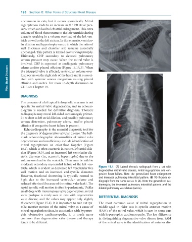
              Ultimately,  CHF  secondary  to  elevated  pulmonary
              venous  pressure  may  occur.  When  the  mitral  valve  is
              involved,  CHF  is  expressed  as  cardiogenic  pulmonary   A
      Misc. Heart Diseases  the tricuspid valve is affected, ventricular volume over-
              edema and/or pleural effusion (Figure 15.1A,B). When

              load occurs on the right side of the heart and it is associ-
              ated  with  systemic  venous  congestion  causing  pleural
              effusion and ascites. For more in-depth discussion on
              CHF, see Chapter 19.


              DIAGNOSIS

              The presence of a left apical holosystolic murmur is not
              specific for mitral valve degeneration, and an echocar-
              diogram  is  needed  for  definitive  diagnosis.  Thoracic
              radiographs may reveal left-sided cardiomegaly primar-
              ily evident as left atrial dilation, and possibly pulmonary
              venous  distension,  pulmonary  edema,  and/or  pleural
              effusion if congestive heart failure is present.
                 Echocardiography is the essential diagnostic tool for
              the diagnosis of degenerative valvular disease. The hall-
              mark  echocardiographic  abnormalities  of  mitral  valve
              degeneration and insufficiency include identification of
              mitral  regurgitation  on  color-flow  Doppler  (Figure
              15.2), which is often eccentric in nature, left atrial dila-
              tion (Figure 15.3), and an increased left ventricular dia-
              stolic diameter (i.e., eccentric hypertrophy) due to the
              volume overload to the ventricle. There may be mild to   B
              moderate secondary myocardial failure of the left ven-
              tricle, which is evident as decreased left ventricular free   Figure  15.1.  (A)	 Lateral	 thoracic	 radiograph	 from	 a	 cat	 with
              wall  motion  and  an  increased  end-systolic  diameter.   degenerative	mitral	valve	disease,	mitral	regurgitation,	and	con-
              However,  fractional  shortening  is  typically  normal  to   gestive	 heart	 failure.	 Note	 the	 generalized	 heart	 enlargement
                                                                 and	increased	pulmonary	interstitial	pattern.	(B)	VD	thoracic	ra-
              high,  due  to  the  increased  ventricular  volume  and   diograph	from	the	same	cat	as	in	(A).	Note	the	generalized	car-
              reduced afterload (because of the mitral valve leak). The   diomegaly,	the	increased	pulmonary	interstitial	pattern,	and	the
              septal systolic wall motion is often hyperdynamic. Unlike   dilated	pulmonary	vasculature	(arrow).
              small dogs with myxomatous valve degeneration, mitral
              valve  prolapse  is  rarely  seen  in  cats  with  degenerative   DIFFERENTIAL DIAGNOSIS
              valve  disease,  and  the  valves  may  appear  only  slightly
              thickened (Figure 15.4). It is important to rule out sys-  The  most  common  cause  of  mitral  regurgitation  in
              tolic anterior motion of the mitral valve as a cause of   middle-aged  to  older  cats  is  systolic  anterior  motion
              mitral regurgitation since, in association with hypertro-  (SAM) of the mitral valve, which is seen in some cats
              phic  obstructive  cardiomyopathy,  it  is  much  more   with hypertrophic cardiomyopathy. The key difference
              common  than  degenerative  valve  disease  and  therapy   in distinguishing degenerative valve disease from SAM
              tends to be different.                             of the mitral valve is the identification of anterior dis-

