Page 194 - Feline Cardiology
P. 194
Chapter 15: Acquired Valvular Disease  197


                                    RV                                                  RV
                                                                                                RA

                                                                                     LV
                               LV
                                                                                              LA


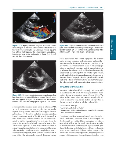
              Figure  15.2.  Right	 parasternal	 long-axis	 color-flow	 Doppler	  Figure 15.4.  Right	parasternal	long-axis	4-chamber	echocardio-
              echocardiogram	of	the	mitral	valve.	Note	that	the	aliased	color-  gram	from	the	same	cat	as	the	previous	images.	Note	the	en-  Misc. Heart Diseases
              flow	pattern	representing	the	high-velocity	jet	of	mitral	regurgita-  larged	left	atrium	(LA)	and	right	atrium	(RA)	and	thickening	of	the
              tion	is	filling	the	left	atrium.	This	echocardiogram	was	obtained	  mitral	valve.	RV	=	right	ventricle;	LV	=	left	ventricle.
              from	the	same	cat	as	the	radiographs	in	Figure	15.1.	LV	=	left
              ventricle;	RV	=	right	ventricle.
                                                                 valve.  Sometimes  with  mitral  dysplasia  the  anterior
                                                                 leaflet appears elongated and misshapen, and papillary
                                                                 muscles may be abnormal in shape and position in the
                                                                 left ventricle. Another common cause of mitral regurgi-
                                                                 tation is functional, secondary mitral regurgitation due
                                                                 to other cardiac diseases such as dilated cardiomyopathy,
                               RA                                unclassified  cardiomyopathy,  or  left-to–right  shunts,
                                     Ao                          which lead to left ventricular enlargement. Secondary, or
                                                                 functional mitral regurgitation, is typically mild or mod-
                                                                 erate and often is symmetrical and centrally arising (in
                                   LA                            the valve orifice), with a normal mitral valve structure.

                                                                 INFECTIVE ENDOCARDITIS

                                                                 Infectious endocarditis (IE) is extremely rare in cats with
                                                                 an incidence of 0.006 to 0.018% of cats presented for exam-
              Figure 15.3.  Right	parasternal	short-axis	echocardiogram	of	the	  ination  in  one  retrospective  report  (Sisson  1994).  The
              heart	base.	Note	the	left	atrial	(LA)	enlargement.	The	right	atrium	  mitral and/or aortic valves are most commonly affected,
              (RA)	 also	 appears	 enlarged.	 This	 echocardigram	 was	 obtained	  which is similar to dogs. Several factors are important in
              from	the	same	cat	as	the	radiographs	in	Figure	15.1.	Ao	=	aorta.  the pathogenesis of infective valvular endocarditis:

                                                                 •  Endothelial damage
              placement of the anterior mitral leaflet in cats with SAM,   •	 Activation	of	clotting	factors
              where  it  approaches  or  touches  the  interventricular   •	 Bacteremia	and	colonization	of	a	noninfective	throm-
              septum during systole. On color-flow Doppler interro-  bus	on	the	valve	surface
              gation, SAM produces two turbulent jets: one ascending
              into the aorta as a result of the left ventricular outflow   Healthy endothelium is not particularly receptive to bac-
              tract obstruction and the other in the left atrium as a   terial  attachment.  However,  when  it  is  damaged,  the
              result  of  mitral  regurgitation.  The  jets  arise  from  the   development  of  platelet-fibrin  aggregates  provides  a
              same point where the valve leaflet causes the obstruction.   susceptible  nidus  for  bacterial  colonization.  Valvular
              Mitral  valve  dysplasia  is  the  most  common  cause  of   endothelium is rich in fibronectin, a glycoprotein pro-
              mitral regurgitation in kittens or juvenile cats. The mitral   duced  following  endothelial  injury.  Many  pathogenic
              valve  typically  has  characteristic  morphologic  abnor-  bacteria  associated  with  IE  have  surface  receptors  for
              malities including short, thick chordae tendinae, which   fibronectin (Peddle and Sleeper 2007), and Staphylococcus
              may  tether  the  abnormally  shaped  thickened  mitral   aureus, which possesses fibronectin receptors, has been

