Page 199 - Feline Cardiology
P. 199
202  Section E: Other Forms of Structural Heart Disease


                                                                 DIFFERENTIAL DIAGNOSIS
                                                                 Echocardiographically,  circumscribed  masses  may  be
                                                                 abscesses,  granulomas,  cysts,  or  neoplasms.  Chemo-
                                                                 dectomas typically are mixed echogenicity masses that
                                                                 may  best  be  identified  between  the  aorta  and  either
                                                                 atrium, or adjacent to the cranial vena cava in 1 case.
                                                                 Myocardial  infiltration  may  be  focal  (Shinohara  et  al.
                                                                 2005) or diffuse (Brummer and Moïse 1989; Liu et al.
                                                                 1989).  Diffuse  infiltration  may  be  mistaken  for
                                                                 hypertrophic  cardiomyopathy  if  attention  is  not  paid
                                                                 to the variable echogenicity that is usually present and
                                                                 the  hypokinesis  (reduced  motion)  of  the  infiltrated
      Misc. Heart Diseases                                       DIAGNOSTIC TESTING
                                                                 myocardium.


                                                                 Cardiac  neoplasia  in  cats  arises  in  three  clinical
                                                                 contexts:

                                                                 1.  A patient shows clinical signs (e.g., respiratory prob-
                                                                   lems) that ultimately are found to be caused by the
                                                                   effects of the neoplasm
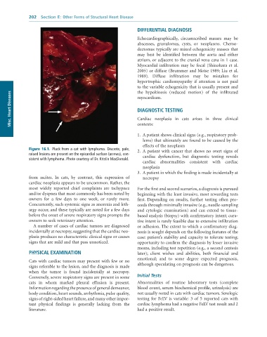
              Figure 16.1.  Pluck	from	a	cat	with	lymphoma.	Discrete,	pale,	  2.  A patient with cancer that shows no overt signs of
              raised	lesions	are	present	on	the	epicardial	surface	(arrows),	con-  cardiac  dysfunction,  but  diagnostic  testing  reveals
              sistent	with	lymphoma.	Photo	courtesy	of	Dr.	Kristin	MacDonald.
                                                                   cardiac  abnormalities  consistent  with  cardiac
                                                                   neoplasia
                                                                 3.  A patient in which the finding is made incidentally at
              from  ascites.  In  cats,  by  contrast,  this  expression  of   necropsy
              cardiac neoplasia appears to be uncommon. Rather, the
              most  widely  reported  chief  complaints  are  tachypnea   For the first and second scenarios, a diagnosis is pursued
              and/or dyspnea that most commonly has been noted by   beginning with the least invasive, most rewarding tests
              owners  for  a  few  days  to  one  week,  or  rarely  more.   first.  Depending  on  results,  further  testing  often  pro-
              Concurrently, such systemic signs as anorexia and leth-  ceeds through minimally invasive (e.g., needle-sampling
              argy occur, and these typically are noted for a few days   and  cytologic  examination)  and  can  extend  to  tissue-
              before the onset of severe respiratory signs prompts the   based analysis (biopsy) with confirmatory intent; cura-
              owners to seek veterinary attention.               tive intent is rarely feasible due to extensive infiltration
                 A number of cases of cardiac tumors are diagnosed   or adhesion. The extent to which a confirmatory diag-
              incidentally at necropsy, suggesting that the cardiac neo-  nosis is sought depends on the following features of the
              plasia produces no characteristic clinical signs or causes   case: patient’s stability and capacity to tolerate testing;
              signs that are mild and that pass unnoticed.       opportunity to confirm the diagnosis by lesser invasive
                                                                 means, including test repetition (e.g., a second centesis
              PHYSICAL EXAMINATION                               later);  client  wishes  and  abilities,  both  financial  and
                                                                 emotional;  and  to  some  degree  expected  prognosis,
              Cats with cardiac tumors may present with few or no
              signs referable to the lesion, and the diagnosis is made   although speculating on prognosis can be dangerous.
              when  the  tumor  is  found  incidentally  at  necropsy.
              Conversely, severe respiratory signs are present in some   Initial Tests
              cats  in  whom  marked  pleural  effusion  is  present.   Abnormalities  of  routine  laboratory  tests  (complete
              Information regarding the presence of general demeanor,   blood count, serum biochemical profile, urinalysis) are
              body condition, heart sounds, arrhythmia, pulse quality,   not usually noted in cats with cardiac tumors. Serologic
              signs of right-sided heart failure, and many other impor-  testing  for  FeLV  is  variable:  3  of  5  reported  cats  with
              tant  physical  findings  is  generally  lacking  from  the   cardiac lymphoma had a negative FeLV test result and 2
              literature.                                        had a positive result.

