
Page 143 - Basic Monitoring in Canine and Feline Emergency Patients
P. 143
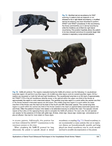
VetBooks.ir                                                Fig. 7.4.  Modified sternal recumbency for FAST
                                                             scanning. In patients that are dyspneic or too
                                                             stressed to be in right lateral recumbency, a modified
                                                             sternal recumbency has been described to perform
                                                                          3
                                                                 3
                                                             AFAST  and TFAST  procedures. In this recumbency,
                                                             the thorax and forelimbs are in sternal recumbency
                                                             while the hindlimbs are shifted into right lateral
                                                             recumbency. This position hopefully allows the patient
                                                             to be less stressed and allows for possible larger tidal
                                                             volumes in respiratory compromised patients.


                                            1

                                              2
                                                      4

                                                 3


             Fig. 7.5.  VetBLUE protocol. The regions evaluated during the VetBLUE protocol are the following: (1) caudodorsal
             lung lobe region; (2) perihilar lung lobe region; (3) middle lung lobe region; and (4) cranial lung lobe region. All four
             regions are assessed on both the left and right hemithorax. The caudodorsal lung lobe is at the level of approximately
             the TFAST  CTS of the left hemithorax (directly dorsal to the xiphoid within the upper one third of the thorax at
                     3
             approximately the eighth to ninth intercostal space). The perihilar lung lobe region is a point within the central one third
             of the thorax between intercostal spaces six and seven. The middle lung lobe region is a point within the lower
             one-third of the thorax over the heart at the level of the fourth and fifth intercostal spaces. The cranial lung lobe
             region is a point within the ventral one third of the thorax cranial to the heart at approximately intercostal spaces two
             and three. Often the patient’s thoracic limb needs to be extended cranially to achieve this view. The operator should
             repeat all four sites on the opposing thorax side to complete the VetBLUE protocol. For each site, the US operator
             can assess for the presence of A-lines with glide sign, B-lines, shred sign, tissue sign, nodule sign, and presence of
             pleural effusion. See text for more detail on these signs.

             in trauma patients. Additionally, this position has   recumbency or standing (Fig. 7.5). Dorsal recumbency is
             not been validated for AFAST  examinations in the   not recommended as many patients that are in respira-
                                    3
             veterinary patient at this time.            tory distress are hemodynamically unstable and being
               When completing the  VetBLUE protocol for lung   placed into dorsal recumbency may be overly stressful
             ultrasound, the patient is typically placed in sternal   and lead to possible decompensation of the patient.


             Applications of Serial Focal Ultrasound Techniques in the Hospitalized Small Animal Patient   135