
Page 35 - Basic Monitoring in Canine and Feline Emergency Patients
P. 35
VetBooks.ir Vena atrium Tricuspid ventricle Pulmonic Pulmonary
Right
Right
valve
valve
artery
Systemic cava Lungs
(pulmonary
circulation
circulation)
Aorta
vein
Left ventricle Mitral Left atrium Pulmonary
Aortic
valve
valve
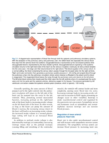
Fig. 2.1. A diagrammatic representation of blood flow through both the systemic and pulmonary circulatory systems.
With the exception of the pulmonary artery and pulmonary vein, the dotted black line separates the venous blood
flow (top) from the arterial blood flow (bottom). Oxygenated blood is represented by the red shaded symbols while
deoxygenated blood is represented by the blue shaded symbols. Deoxygenated blood originating in the systemic
circulation returns to the right-hand side of the heart via the venous circulation. Eventually all venous blood empties
into the vena cava – the large vein that returns blood to the right atrium. This blood equates to the venous return. In
a normal setting, the blood comprising the venous return passes through the tricuspid valve into the right ventricle.
Right ventricular contraction then generates a pulmonary systolic pressure of ~25 mmHg and propels blood through
the pulmonary artery into the pulmonary circulation where carbon dioxide is offloaded to the alveoli and the blood
is oxygenated. This oxygenated blood leaves the lungs through the pulmonary veins and returns to the left atrium.
The left atrial blood volume then travels past the mitral valve into the left ventricle where it is pumped past the aortic
valve and into the aorta and the systemic arteries, delivering oxygen to the tissues. The pressure generated by left
ventricular contraction creates the systemic arterial systolic pressure, which is typically ~120 mmHg.
Generally speaking, the same amount of blood muscle), the ventricle will contract harder and more
pumped out by the right ventricle into the pulmo- completely, ejecting more blood into the aorta.
nary circulation will return to the left side of the Increasing inotropy leads to increasing stroke vol-
heart and be pumped into the aorta by the left ume and an increase in MAP (again, presuming no
ventricle. Thus, presuming normal myocardial changes occur in other variables). Inotropy is
function, increasing preload on the right-hand under neurohormonal control and regulated by
side of the heart leads to increasing stroke volume the autonomic nervous system. Sympathetic nerves
on the left-hand side of the heart. In other words, and hormones (such as epinephrine and norepi-
as long as the heart can pump the amount of nephrine) increase inotropy and can increase
blood it receives, the increasing venous return to blood pressure.
the heart will lead to an increasing stroke volume.
An increased stroke volume in a normal physio-
logic setting will lead to an increased blood Regulation of mean arterial
pressure. pressure: Heart rate
In addition to preload, stroke volume is also Heart rate is also under neurohormonal control
determined by inotropy (or contractility). Increasing and will increase with sympathetic nervous system
inotropy means that as the preload increases stimulation. The sympathetic nervous system can
(causing filling and stretching of the ventricular increase blood pressure by increasing heart rate
Blood Pressure Monitoring 27