
Page 1154 - Veterinary Toxicology, Basic and Clinical Principles, 3rd Edition
P. 1154
1086 SECTION | XVI Feed and Water Contaminants
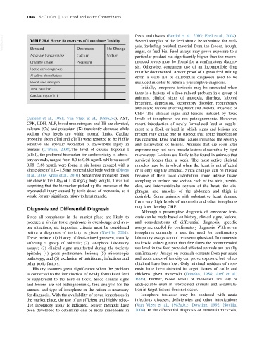
VetBooks.ir TABLE 78.6 Some Biomarkers of Ionophore Toxicity feeds and tissues (Bertini et al., 2003; Ebel et al., 2004).
Several samples of the feed should be submitted for anal-
ysis, including residual material from the feeder, trough,
Elevated
Decreased
No Change
auger, or feed bin. Feed assays may prove exposure to a
Aspartate transaminase Calcium Sodium particular product but significantly higher than the recom-
Creatine kinase Potassium mended levels must be found for a confirmatory diagno-
sis. Otherwise, concurrent use of an incompatible drug
Lactic dehydrogenase
must be documented. Absent proof of a gross feed mixing
Alkaline phosphatase
error, a wide list of differential diagnoses need to be
Blood urea nitrogen excluded in order to return a presumptive diagnosis.
Initially, ionophore toxicosis may be suspected when
Total bilirubin
there is a history of a feed-related problem in a group of
Cardiac troponin 1
animals; clinical signs of anorexia, diarrhea, labored
breathing, depression, locomotory disorder, recumbency
and death; lesions affecting heart and skeletal muscles; or
CHF. The clinical signs and lesions induced by toxic
(Amend et al., 1981; Van Vleet et al., 1983a,b,c). AST, levels of ionophores are not pathognomonic. However,
CPK, LDH, ALP, blood urea nitrogen, and TB are elevated, recent introduction of newly formulated feed or supple-
calcium (Ca) and potassium (K) transiently decrease while ment to a flock or herd in which signs and lesions are
sodium (Na) levels are within normal limits. Cardiac present may cause one to suspect that acute intoxication
troponins (both cTnI and cTnT) were reported to be highly has occurred. Dose and time factors influence the severity
sensitive and specific biomarker of myocardial injury in and distribution of lesions. Animals that die soon after
humans (O’Brien, 2008).The level of cardiac troponin I exposure may not have muscle lesions discernible by light
(cTnI), the preferred biomarker for cardiotoxicity in labora- microscopy. Lesions are likely to be found in animals that
tory animals, ranged from 0.0 to 0.06 ng/mL while values of survived longer than a week. The most active skeletal
0.08 3.68 ng/mL were found in six horses gavaged with a muscles may be involved when the heart is not affected
single dose of 1.0 1.5 mg monensin/kg body weight (Divers or is only slightly affected. Since changes can be missed
et al., 2009; Kraus et al., 2010). Since these monensin doses because of their focal distribution, more intense tissue
are close to the LD 50 of 1.38 mg/kg body weight, it was not sampling to include one section each of the atria, ventri-
surprising that the biomarker picked up the presence of the cles, and interventricular septum of the heart, the dia-
myocardial injury caused by toxic doses of monensin, as it phragm, and muscles of the abdomen and thigh is
would for any significant injury to heart muscle. desirable. Some animals with substantive heart damage
from very high levels of monensin and other ionophores
may later develop CHF.
Diagnosis and Differential Diagnosis
Although a presumptive diagnosis of ionophore toxi-
Since all ionophores in the market place are likely to cosis can be made based on history, clinical signs, lesions,
produce a similar toxic syndrome in overdosage and mis- and considerations of differential diagnosis, specific
use situations, six important criteria must be considered assays are needed for confirmatory diagnosis. With seven
before a diagnosis of toxicity is given (Novilla, 2004). ionophores currently in use, the need for confirmatory
These include (1) history of feed-related problem, usually laboratory assays cannot be overemphasized. In monensin
affecting a group of animals; (2) ionophore laboratory toxicosis, values greater than five times the recommended
assays; (3) clinical signs manifested during the toxicity use level in the feed provided affected animals are usually
episode; (4) gross postmortem lesions; (5) microscopic confirmatory. Assays on stomach contents from per acute
pathology; and (6) exclusion of nutritional, infectious and and acute cases of toxicity can prove exposure but values
other toxic factors. obtained have been low. Only minimal residues of mon-
History assumes great significance when the problem ensin have been detected in target tissues of cattle and
is connected to the introduction of newly formulated feed chickens given monensin (Donoho, 1984; Atef et al.,
or supplement to the herd or flock. Since clinical signs 1993). Further, blood levels of monensin are low or
and lesions are not pathognomonic, feed analysis for the undetectable even in intoxicated animals and accumula-
amount and type of ionophore in the ration is necessary tion in target tissues does not occur.
for diagnosis. With the availability of seven ionophores in Ionophore toxicosis may be confused with acute
the market place, the use of an efficient and highly selec- infectious diseases, deficiencies and other intoxications
tive laboratory assay is indicated. Newer methods have (Van Vleet et al., 1983a,b,c; Dowling, 1992; Novilla,
been developed to determine one or more ionophores in 2004). In the differential diagnosis of monensin toxicosis,