
Page 1277 - Small Animal Internal Medicine, 6th Edition
P. 1277
CHAPTER 73 Common Immune-Mediated Diseases 1249
other adjunctive immunosuppressive drugs (e.g., azathio- the glomerular capillary wall by a carbohydrate-glycoprotein
prine, cyclosporine, mycophenolate) is usually necessary to interaction.
VetBooks.ir induce or maintain remission. Little information is avail- consequences are similar (see Chapter 40) and ultimately
Whatever the cause of immune complex deposition, the
able on the efficacy of drug protocols for treating SLE. The
prognosis for dogs with SLE is guarded to poor. Relapse is
failure, and predisposition to thromboembolism.
common regardless of the drug protocol used, and long- lead to severe proteinuria, systemic hypertension, renal
term and often lifelong immunosuppressive therapy is
necessary to control the disease. Relapses may involve dif- Clinical Features
ferent organ systems and clinical signs than at initial pre- The hallmark of GN is proteinuria, which is readily detected
sentation (e.g., hemolytic anemia initially and polyarthritis on routine urinalysis. In many cases proteinuria is initially
at relapse). identified as an incidental finding, and the animal may have
no obvious clinical signs or only subtle abnormalities (e.g.,
weight loss, lethargy, decreased appetite). In other cases,
GLOMERULONEPHRITIS animals present with clinical signs of renal failure (e.g.,
anorexia, weight loss, vomiting, polyuria, polydipsia), and
Etiology proteinuria is identified in the course of the evaluation. In
Acquired GN is more common in dogs than cats and results nephrotic syndrome, which is defined as the presence of
from the presence of immune complexes within the glo- proteinuria, hypoalbuminemia, hypercholesterolemia, and
merular capillary walls (see Chapter 40). Immune com- either peripheral edema or ascites, the clinical signs are more
plexes may be circulating antigen-antibody complexes that severe and often rapidly progressive. Other clinical signs in
are deposited or trapped in the glomerulus or may form in dogs with GN may relate to the presence of hypertension
situ when circulating antibodies react with either endoge- or hypercoagulability. Hypertension may result in retinal
nous glomerular antigens or nonglomerular antigens within changes and blindness, whereas TEs may occur as a result of
the glomerular capillary wall. Soluble circulating immune the hypercoagulable state.
complexes formed in the presence of mild antigen excess,
or when both antigen and antibody are present in approxi- Diagnosis
mately equal quantities, may be deposited along capillary A diagnosis of protein-losing nephropathy (PLN) is made
walls, resulting in a granular pattern observed on immuno- by documentation of persistent proteinuria that cannot be
fluorescent or immunoperoxidase staining. Infectious and explained by inflammation of the lower urinary tract or
inflammatory diseases are common identifiable causes for blood contamination of the urine. Initial dipstick estimates
deposition of immune complexes within the glomerulus of urine protein should be evaluated in the light of the urine
(Box 73.6). Unfortunately, in the majority of cases of GN, an sediment and specific gravity of the urine. The severity of
underlying cause is not identified. When immune complexes protein loss should then be quantitated by measurement of
form in situ, a smooth linear pattern is observed with immu- a protein-to-creatinine ratio, preferably on a urine sample
nofluorescent or immunoperoxidase staining. Causes of in with no inflammation or hematuria. A protein-to-creatinine
situ deposition of immune complexes may be either true ratio greater than 0.5 is abnormal; most dogs and cats with
autoimmune disease when antibodies are directed against PLN have a ratio greater than 2.0. Once persistent protein-
the basement membrane of the glomerular capillaries or uria has been documented, further testing is necessary to
when antigen becomes localized in the glomerular capillary determine whether evidence of tubular dysfunction also
wall. For example, in dogs with heartworm disease, soluble exists and to investigate for the presence of underlying infec-
Dirofilaria immitis antigens have been shown to adhere to tious or inflammatory diseases implicated as causes of GN.
Diagnostic tests that should be performed include a CBC,
serum biochemical profile, urinalysis and urine culture,
blood pressure, and radiographs of the thorax and abdomen.
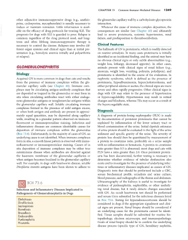
BOX 73.6 Ultrasonography of the kidneys is useful to investigate for
evidence of pyelonephritis, nephroliths, or other underly-
Infectious and Inflammatory Diseases Implicated in ing renal disease, but it rarely detects changes associated
Pathogenesis of Glomerulonephritis in Dogs with GN. An occult heartworm test should be performed
and serum titers submitted for the infectious diseases listed
Ehrlichiosis in Box 73.6. Testing for hyperadrenocorticism should be
Dirofilariasis considered in dogs if the appropriate signalment and clini-
Leptospirosis cal signs are present. Renal biopsy should be considered if
Borreliosis
Brucellosis an underlying cause for the proteinuria cannot be identi-
Endocarditis fied. Tissue samples should be submitted for routine his-
Pyelonephritis topathology, electron microscopy, and immunopathology.
Prostatitis Goals of renal biopsy should be to confirm the underlying
disease process (specific type of GN, hereditary nephritis,